Vesicoureteral Reflux on Radionuclide Cystogram
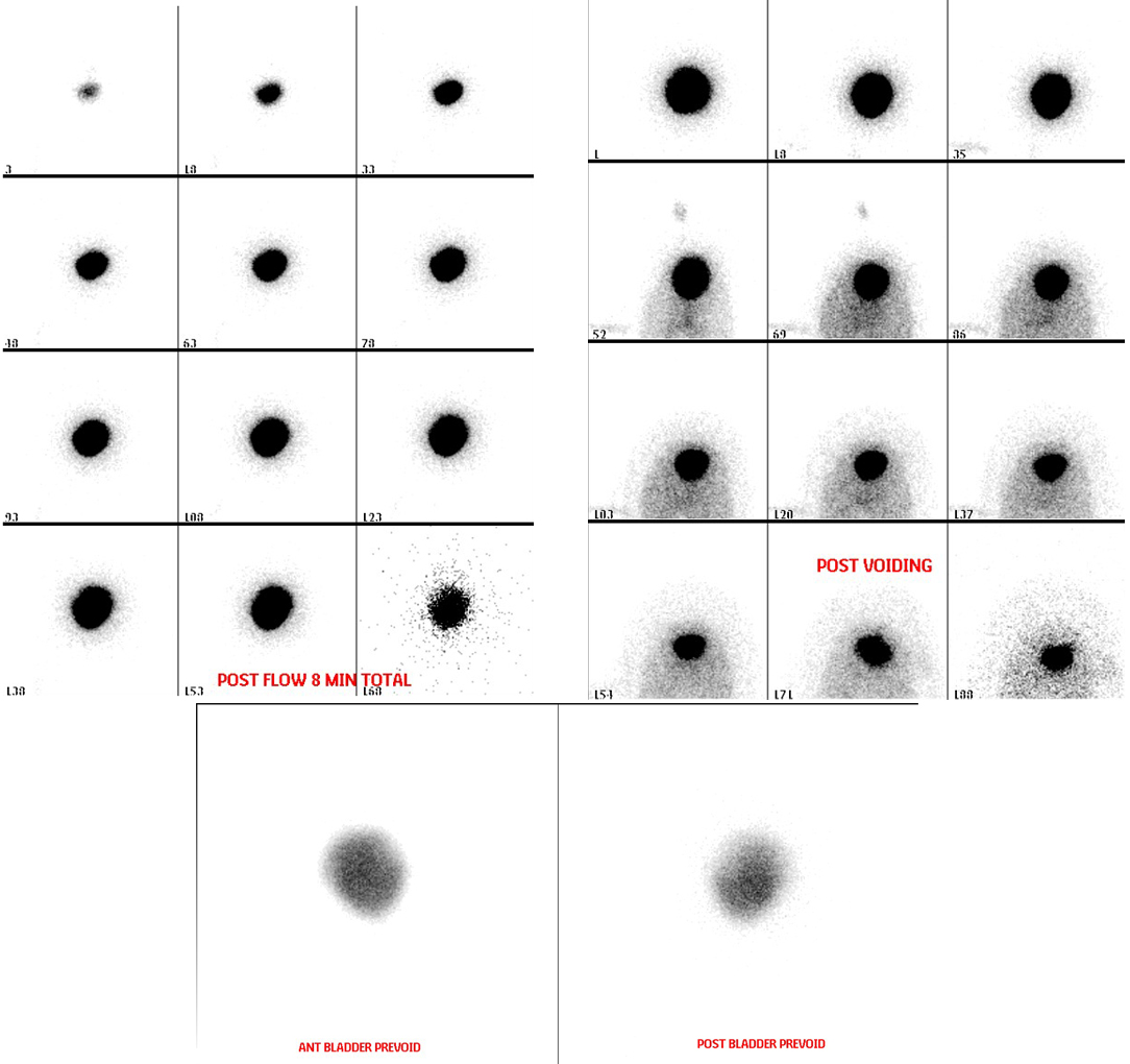
Provided clinical image
Question
During a direct radionuclide cystogram like the one shown, what specific finding on the images indicates that VESICOURETERAL REFLUX (VUR) is present?
- AThe bladder fails to empty completely on post-voiding images, leaving significant residual tracer activity in the bladder after the patient voids
- BRadiotracer activity appears in the ureters or kidneys during bladder filling or voiding, indicating urine flowing backward from the bladder toward the kidneys
- CThe bladder appears enlarged and asymmetric on pre-void images, indicating increased bladder capacity due to detrusor muscle hypertrophy
- DActivity is seen only in the urethra during voiding, indicating normal antegrade urine flow without any evidence of retrograde reflux
Show the answer and explanation
B. Radiotracer activity appears in the ureters or kidneys during bladder filling or voiding, indicating urine flowing backward from the bladder toward the kidneys
Vesicoureteral reflux (VUR) is identified when radiotracer activity is seen moving from the bladder upward into one or both ureters and/or renal pelves during the filling or voiding phase. Normally, tracer should remain confined to the bladder during filling and exit only through the urethra during voiding. Any appearance of tracer in the upper urinary tract (ureters or kidneys) is abnormal and indicates retrograde flow — VUR. The direct method is very sensitive for VUR because imaging is continuous throughout the entire filling and voiding cycle, catching both low-pressure and high-pressure reflux.
More renal and genitourinary images
Why Post-Void Images on a Radionuclide Cystogram
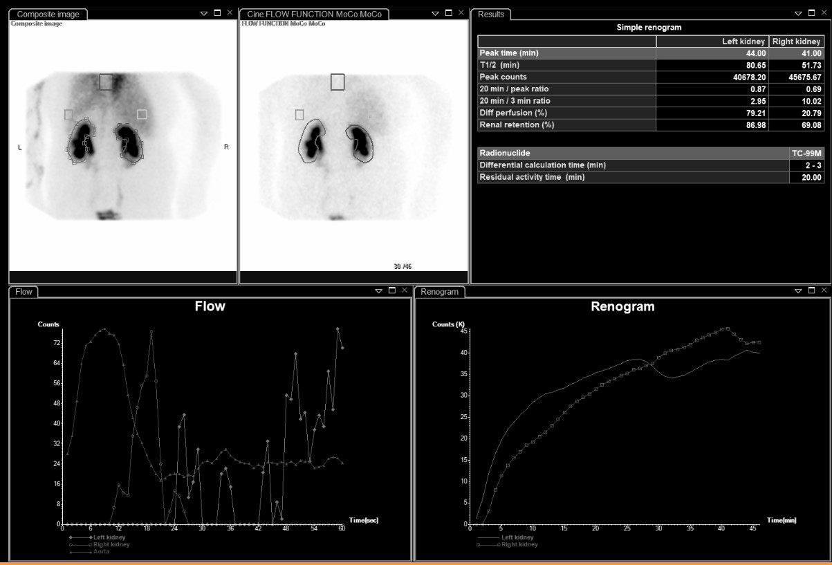 Differential Renal Function on MAG3
Bilateral Impaired Drainage on MAG3 Renogram
Motion Correction in MAG3 Renal Scans
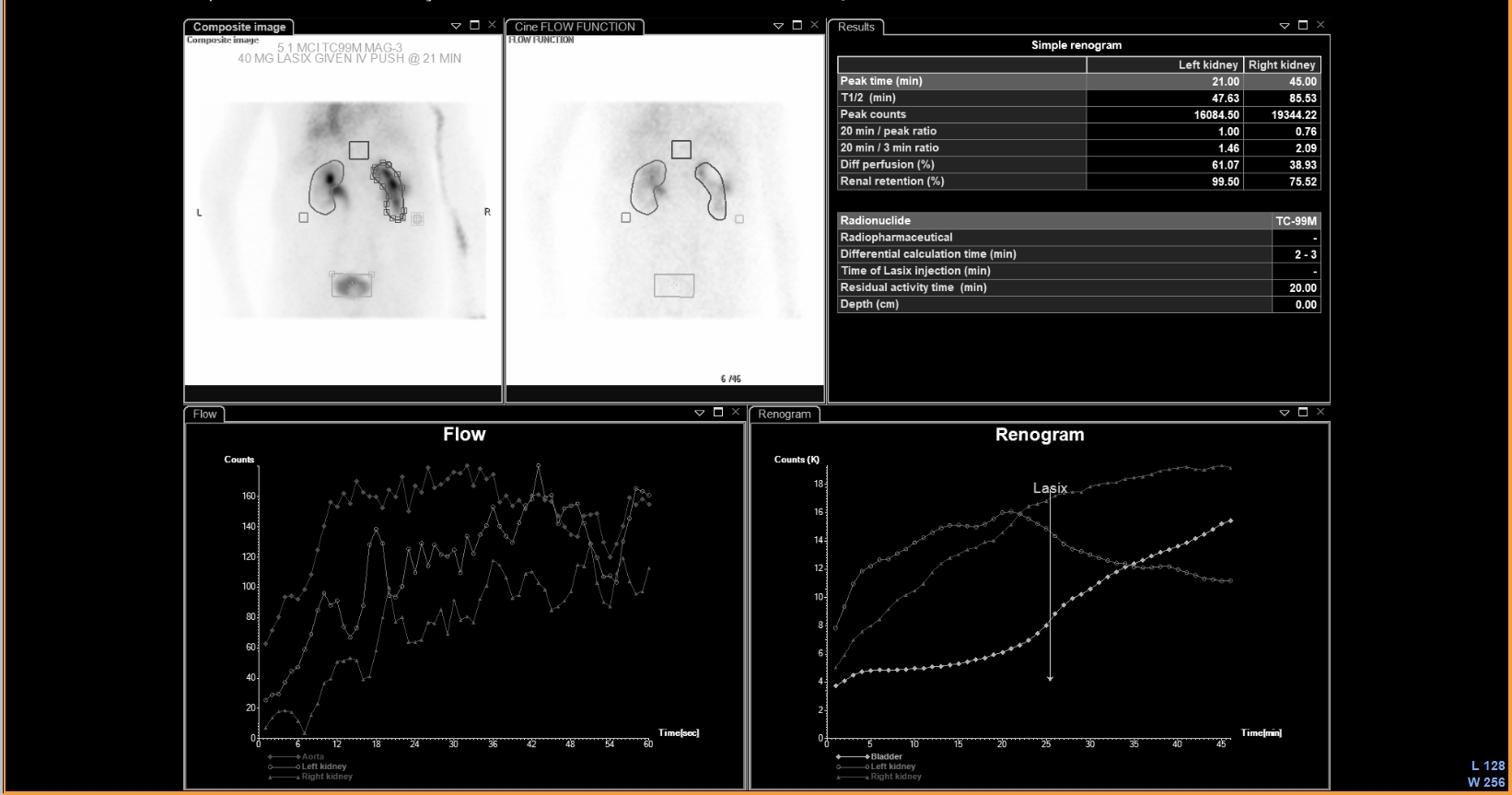 UPJ Obstruction on MAG3 Diuretic Renogram
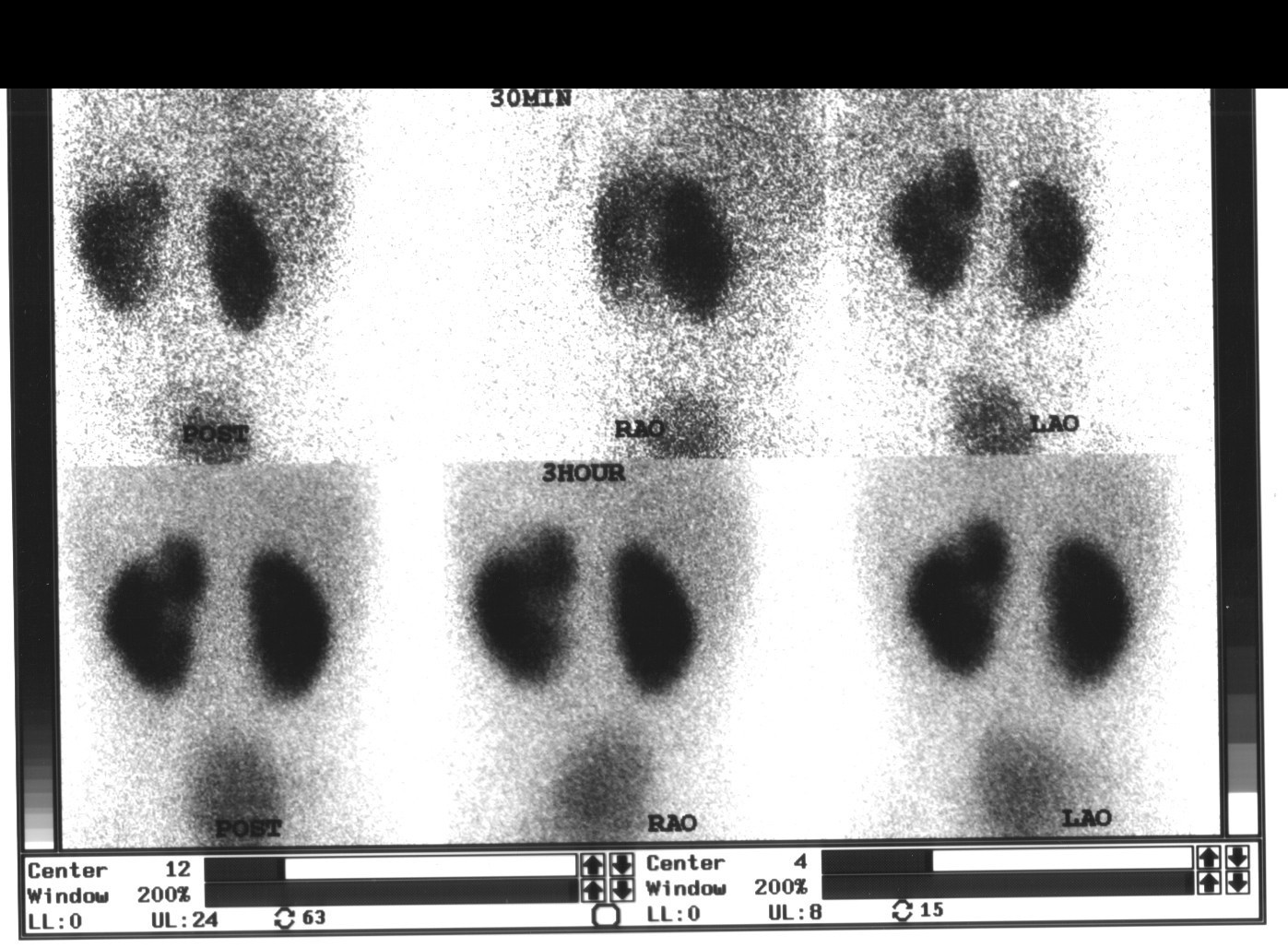 Renal Cortical Scar on DMSA Scan
All 130 nuclear medicine images by category →
Practise image interpretation in the app
All 130 images as an interactive quiz, alongside 500 NMTCB and ARRT(N) questions with worked explanations, case studies and timed mock exams. 25 questions are free, no card and no account.