Renal Cortical Scar on DMSA Scan
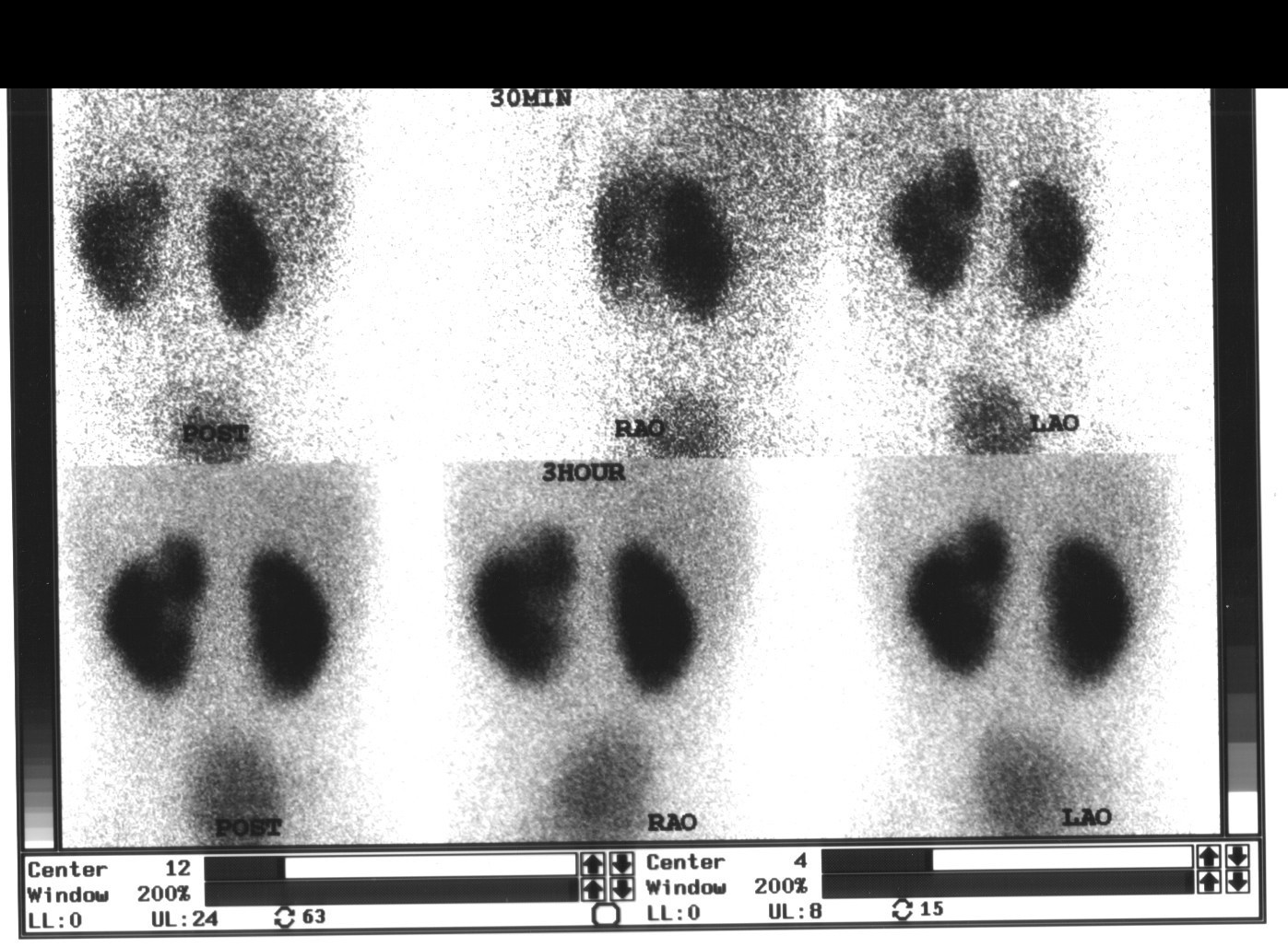
Provided clinical image
Question
A Tc-99m DMSA (dimercaptosuccinic acid) renal cortical scan is performed in a child after a urinary tract infection. The scan shows a focal area of decreased uptake in the upper pole of the left kidney. This finding is MOST consistent with:
- ALeft renal artery stenosis causing upper pole ischemia
- BRenal cortical scar (pyelonephritic scar) from prior pyelonephritis — DMSA is the gold standard for detecting cortical scarring
- CWilms' tumor (nephroblastoma)
- DHydronephrosis
Show the answer and explanation
B. Renal cortical scar (pyelonephritic scar) from prior pyelonephritis — DMSA is the gold standard for detecting cortical scarring
Tc-99m DMSA is the gold standard for detecting renal cortical scarring after pyelonephritis. DMSA is taken up by functioning renal tubular cells and retained in the cortex, so areas of cortical scar (replaced by fibrous tissue with non-functioning tubules) appear as focal photopenic defects. In children, UTIs can cause pyelonephritis and subsequent cortical scarring leading to hypertension and chronic kidney disease — making DMSA scanning essential for follow-up after febrile UTI. DMSA can also detect acute pyelonephritis (areas of decreased uptake that may recover). Sensitivity for cortical scar is >90%. DMSA also measures differential (split) renal function.
More renal and genitourinary images
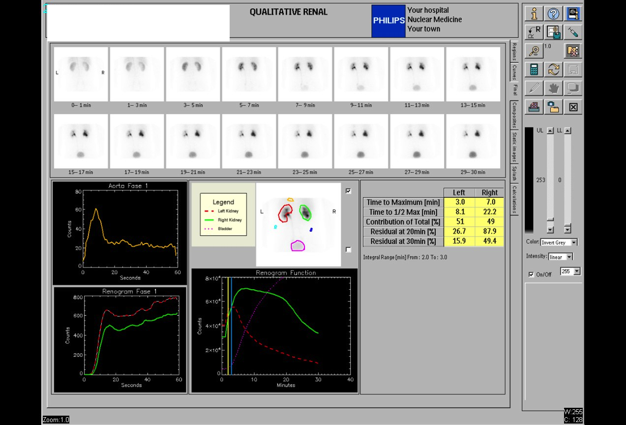 Normal Renal Drainage Half-Time on MAG3
Why MAG3 for Renal Imaging: Tubular Secretion
Furosemide Dose for Diuretic Renography
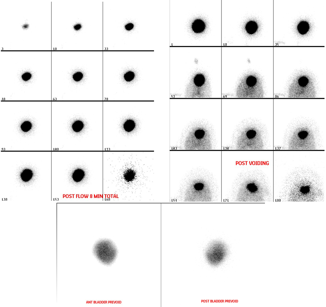 Direct Radionuclide Cystogram Technique
Vesicoureteral Reflux on Radionuclide Cystogram
Why Post-Void Images on a Radionuclide Cystogram
All 130 nuclear medicine images by category →
Practise image interpretation in the app
All 130 images as an interactive quiz, alongside 500 NMTCB and ARRT(N) questions with worked explanations, case studies and timed mock exams. 25 questions are free, no card and no account.