UPJ Obstruction on MAG3 Diuretic Renogram
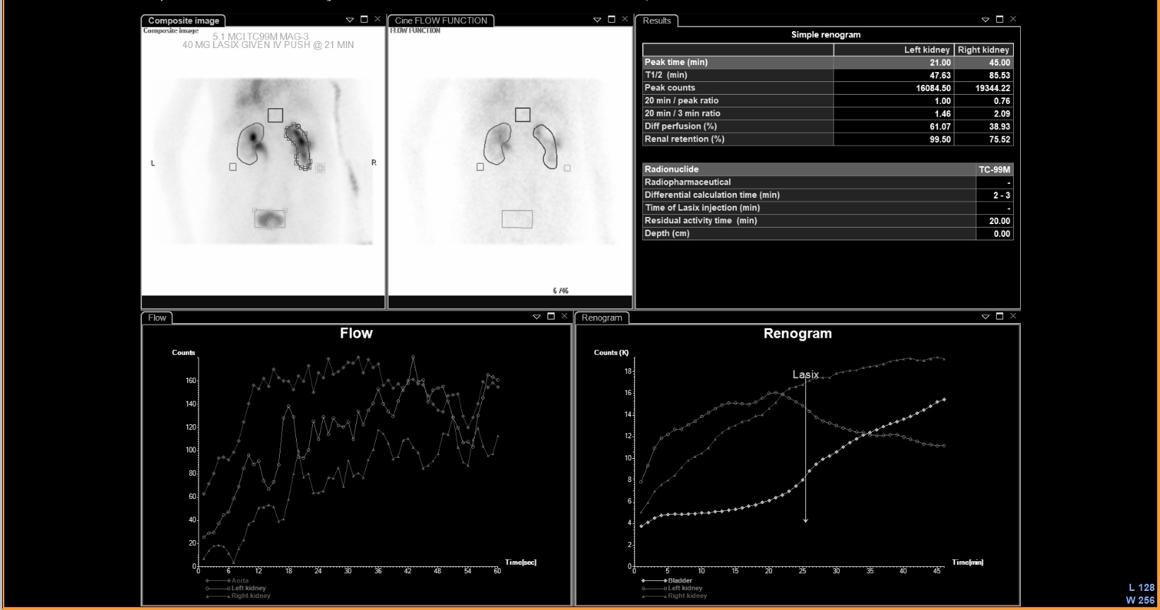
Provided clinical image
Question
A Tc-99m MAG3 (mercaptoacetyltriglycine) diuretic renogram is performed to evaluate a dilated right renal collecting system (hydronephrosis) found on ultrasound. The MAG3 scan shows normal tracer uptake by both kidneys, but tracer is retained in the right renal pelvis after furosemide (Lasix) injection and shows a prolonged T½ of 28 minutes (normal <10 min). This is MOST consistent with:
- AObstructed right ureteropelvic junction (UPJ) — prolonged T½ after diuretic confirms true mechanical obstruction
- BNormal variant dilated collecting system — no obstruction
- CRenal artery stenosis
- DVesicoureteral reflux
Show the answer and explanation
A. Obstructed right ureteropelvic junction (UPJ) — prolonged T½ after diuretic confirms true mechanical obstruction
Tc-99m MAG3 diuretic renography (Lasix renogram) differentiates obstructed from non-obstructed dilated collecting systems. After IV furosemide, a normal or dilated-but-not-obstructed system drains rapidly (T½ < 10 min). In true mechanical obstruction (UPJ obstruction, ureteral stricture), the tracer cannot drain despite diuretic washout and T½ is markedly prolonged (>20 min = obstructed; 10–20 min = equivocal). A T½ of 28 minutes confirms right UPJ obstruction requiring urologic intervention (pyeloplasty). Tc-99m MAG3 is preferred for renal function assessment as it is tubularly secreted (not filtered like DTPA), providing better image quality even in impaired renal function.
More renal and genitourinary images
 Renal Cortical Scar on DMSA Scan
 Normal Renal Drainage Half-Time on MAG3
Why MAG3 for Renal Imaging: Tubular Secretion
Furosemide Dose for Diuretic Renography
 Direct Radionuclide Cystogram Technique
Vesicoureteral Reflux on Radionuclide Cystogram
All 130 nuclear medicine images by category →
Practise image interpretation in the app
All 130 images as an interactive quiz, alongside 500 NMTCB and ARRT(N) questions with worked explanations, case studies and timed mock exams. 25 questions are free, no card and no account.