Direct Radionuclide Cystogram Technique
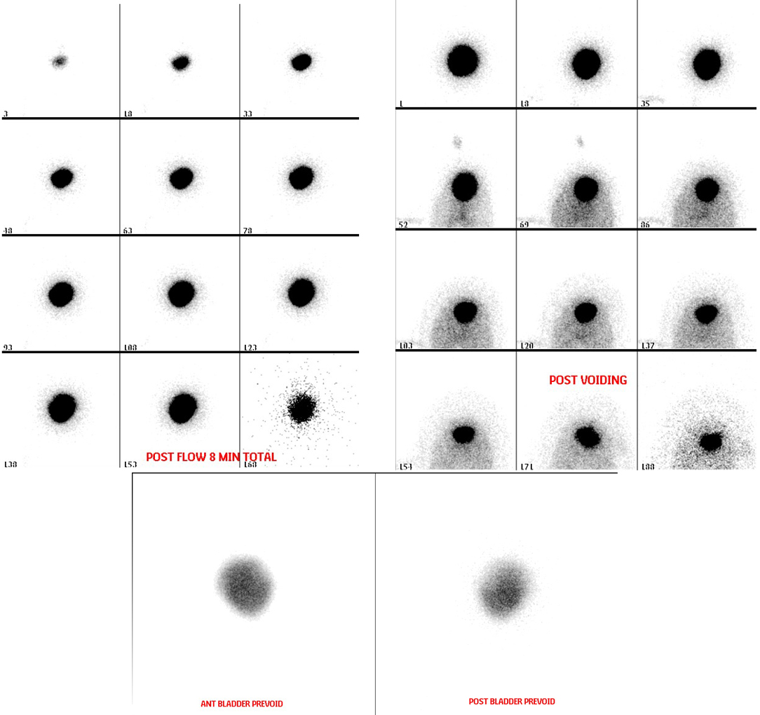
Provided clinical image
Question
The images shown are from a direct radionuclide cystogram (DRC). As the nuclear medicine technologist performing this procedure, how is the radiotracer introduced into the patient?
- AThe tracer is injected intravenously — it is filtered by the kidneys and travels down the ureters to fill the bladder naturally over 30–60 minutes (indirect method)
- BThe patient drinks a dilute oral solution of the tracer, which is absorbed by the gastrointestinal tract and concentrated in the urine within 2–3 hours
- CThe tracer is injected subcutaneously in the lower abdomen, where it is absorbed by the lymphatic system and drains into the bladder via the pelvic lymphatics
- DThe tracer is instilled directly into the bladder through a urinary catheter, mixed with sterile saline, which fills the bladder while imaging is performed
Show the answer and explanation
D. The tracer is instilled directly into the bladder through a urinary catheter, mixed with sterile saline, which fills the bladder while imaging is performed
In direct radionuclide cystography (DRC), a urinary catheter is inserted and a small amount of Tc-99m pertechnetate (or Tc-99m sulfur colloid) diluted in sterile saline is instilled directly into the bladder. Images are acquired continuously during filling and voiding to detect vesicoureteral reflux (VUR). This differs from INDIRECT cystography, where tracer is given IV and imaged after it fills the bladder through the kidneys — no catheter is needed for indirect, but it cannot detect reflux during the filling phase.
More renal and genitourinary images
Vesicoureteral Reflux on Radionuclide Cystogram
Why Post-Void Images on a Radionuclide Cystogram
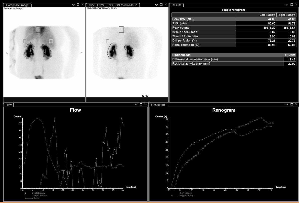 Differential Renal Function on MAG3
Bilateral Impaired Drainage on MAG3 Renogram
Motion Correction in MAG3 Renal Scans
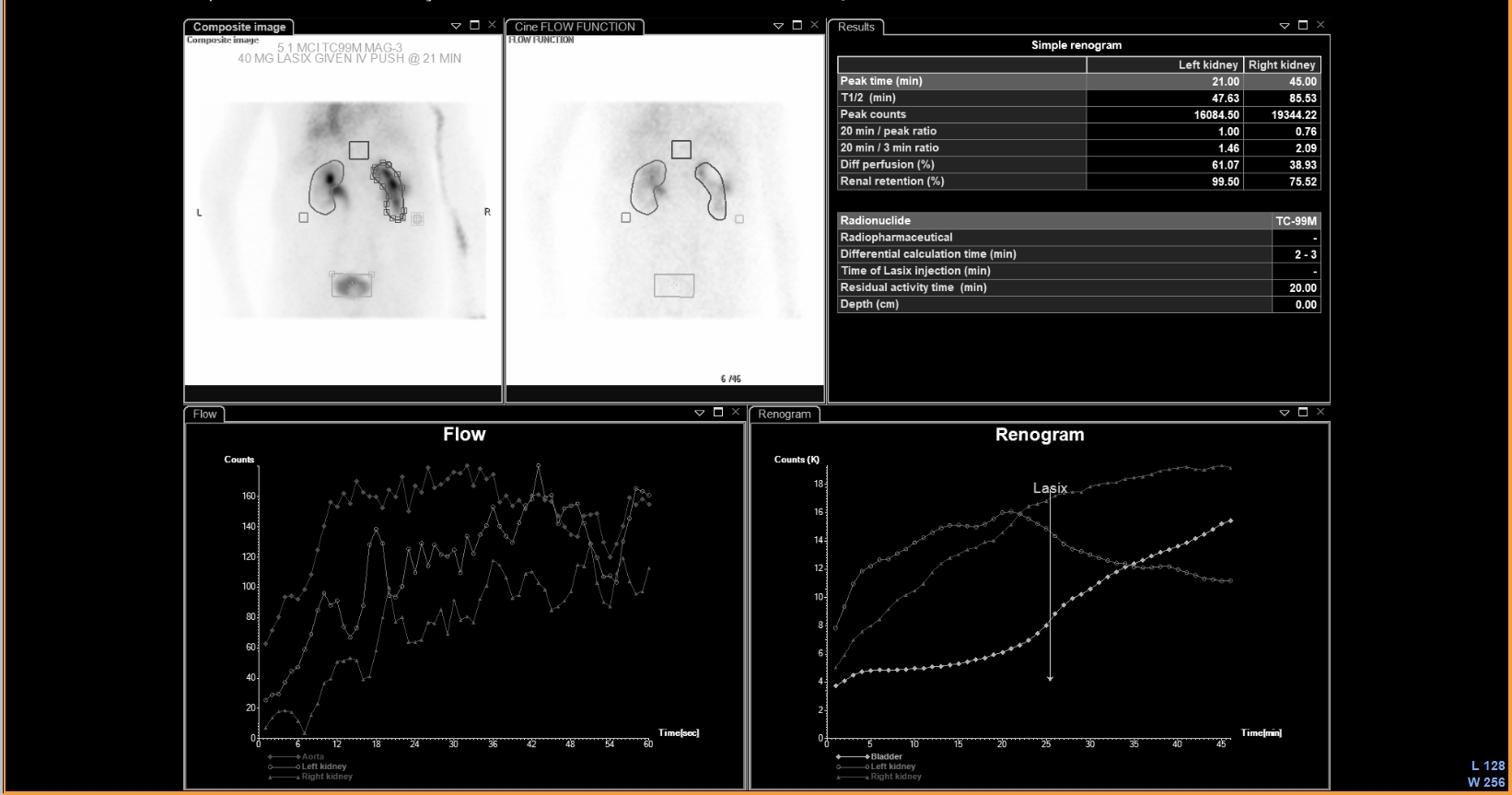 UPJ Obstruction on MAG3 Diuretic Renogram
All 130 nuclear medicine images by category →
Practise image interpretation in the app
All 130 images as an interactive quiz, alongside 500 NMTCB and ARRT(N) questions with worked explanations, case studies and timed mock exams. 25 questions are free, no card and no account.