Ventilation Agents: Tc-99m DTPA Aerosol vs Xe-133
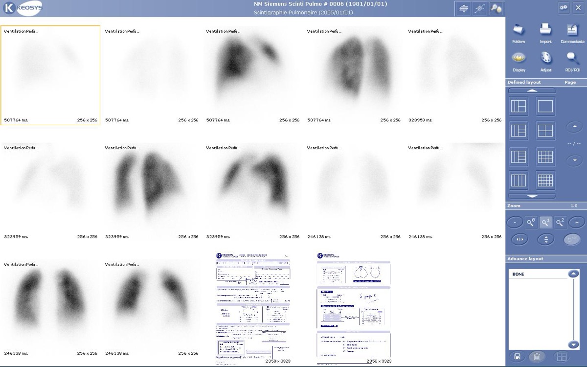
Public domain · Wikimedia Commons · source file
Question
Two ventilation agents are used for V/Q lung scanning: Xe-133 (xenon gas) and Tc-99m DTPA aerosol. Which of the following correctly compares these two agents?
- ATc-99m DTPA aerosol can be imaged in multiple projections; Xe-133 is limited to a single-detector projection due to its poor 81 keV photon energy
- BXe-133 provides better spatial resolution than Tc-99m aerosol
- CTc-99m DTPA aerosol requires a gas-handling exhaust system
- DXe-133 and Tc-99m aerosol are equivalent in all clinical situations
Show the answer and explanation
A. Tc-99m DTPA aerosol can be imaged in multiple projections; Xe-133 is limited to a single-detector projection due to its poor 81 keV photon energy
Xe-133 (81 keV gamma, 5.3-day half-life) and Tc-99m DTPA aerosol (140 keV) differ importantly: (1) Xe-133 is imaged at a SINGLE projection (usually posterior) because the patient must breathe the gas continuously and cannot be repositioned; it requires a gas-handling system with exhaust. (2) Tc-99m DTPA aerosol is inhaled as an aerosol and deposits in the lungs, after which the patient can be repositioned for multiple planar projections or SPECT — dramatically improving diagnostic quality. (3) Tc-99m aerosol has better photon energy (140 keV) for gamma camera imaging. However, in central airway disease (COPD), aerosol may clump in the central airways ('hot spots'), while Xe-133 distributes more uniformly.
More lung v/q imaging images
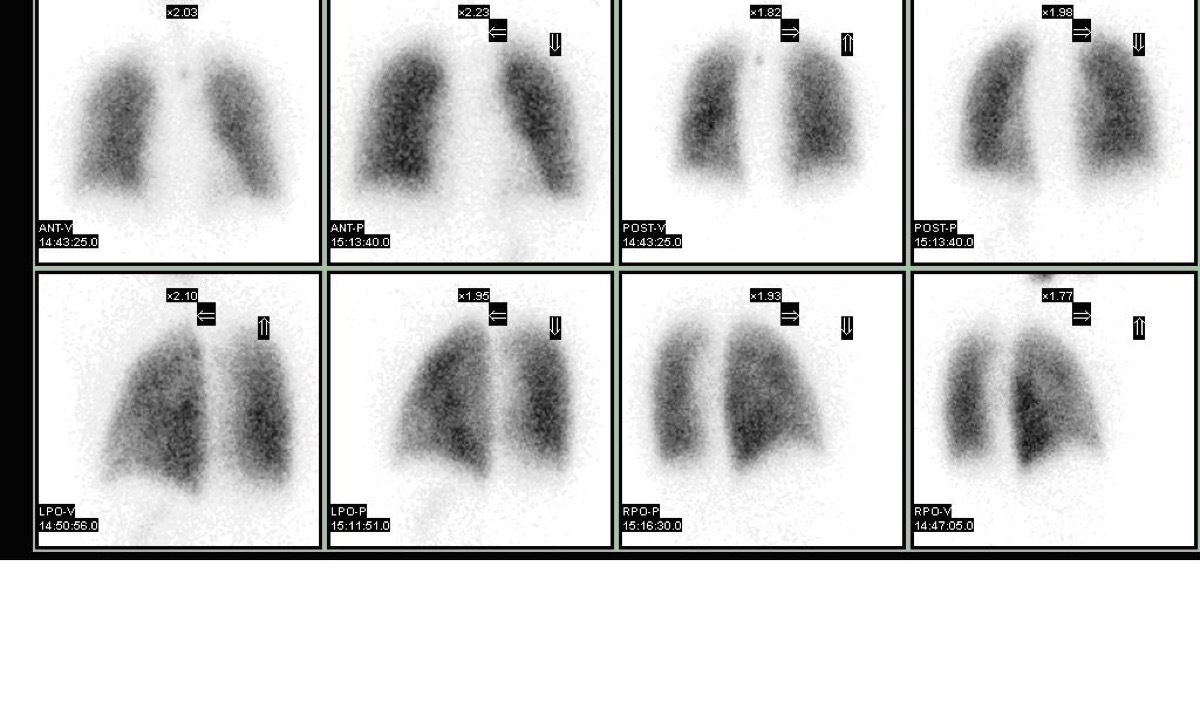 Intermediate-Probability V/Q Scan
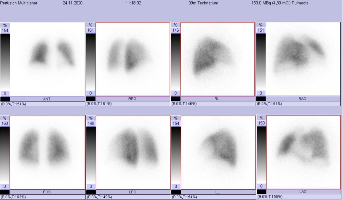 Brain and Kidney MAA Uptake: Right-to-Left Shunt
Tc-99m MAA Injection Technique
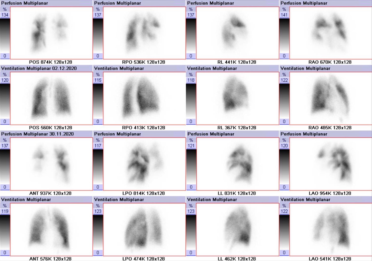 PIOPED II: High-Probability V/Q with High Pretest Probability
Wedge-Shaped Pleural-Based Defect: Pulmonary Embolism
Lung Perfusion Projections: Why Lateral Views Matter
All 130 nuclear medicine images by category →
Practise image interpretation in the app
All 130 images as an interactive quiz, alongside 500 NMTCB and ARRT(N) questions with worked explanations, case studies and timed mock exams. 25 questions are free, no card and no account.