Wedge-Shaped Pleural-Based Defect: Pulmonary Embolism
CC BY 3.0 · Myohan / Wikimedia Commons · source file
Question
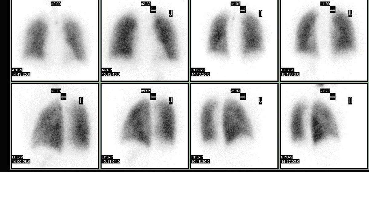
On a lung perfusion scan, a wedge-shaped, PLEURAL-BASED perfusion defect (broad base at the pleural surface, apex pointing toward the hilum) is identified. The ventilation scan is normal in the same region. This WEDGE-SHAPED peripheral mismatch is the classic scintigraphic appearance of:
- APleural effusion
- BPulmonary contusion
- CPneumonia with consolidation
- DPulmonary embolism — embolic occlusion of a segmental or subsegmental pulmonary artery
Show the answer and explanation
D. Pulmonary embolism — embolic occlusion of a segmental or subsegmental pulmonary artery
The classic scintigraphic appearance of PE on perfusion scan is a WEDGE-SHAPED (or 'pleural-based') perfusion defect, where the broad base is at the pleural surface and the narrow apex points toward the hilum — following the anatomic distribution of a pulmonary artery branch. This shape reflects the vascular anatomy of a segmental or subsegmental artery territory. The corresponding ventilation scan is NORMAL (ventilation-perfusion mismatch). Multiple such wedge-shaped defects in different lung zones constitute a high probability pattern. Pleural effusion causes a uniformly decreased peripheral perfusion without a discrete wedge shape.
More lung v/q imaging images
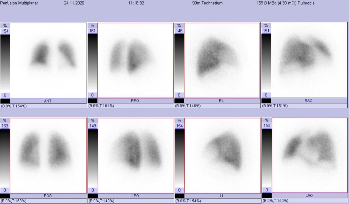 Lung Perfusion Projections: Why Lateral Views Matter
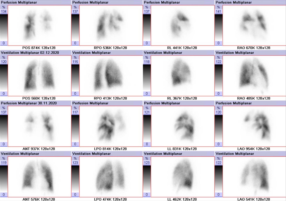 High-Probability V/Q Scan: Mismatched Defects
Matched V/Q Defects: Low Probability for PE
Normal Lung Perfusion Scan and Pulmonary Embolism
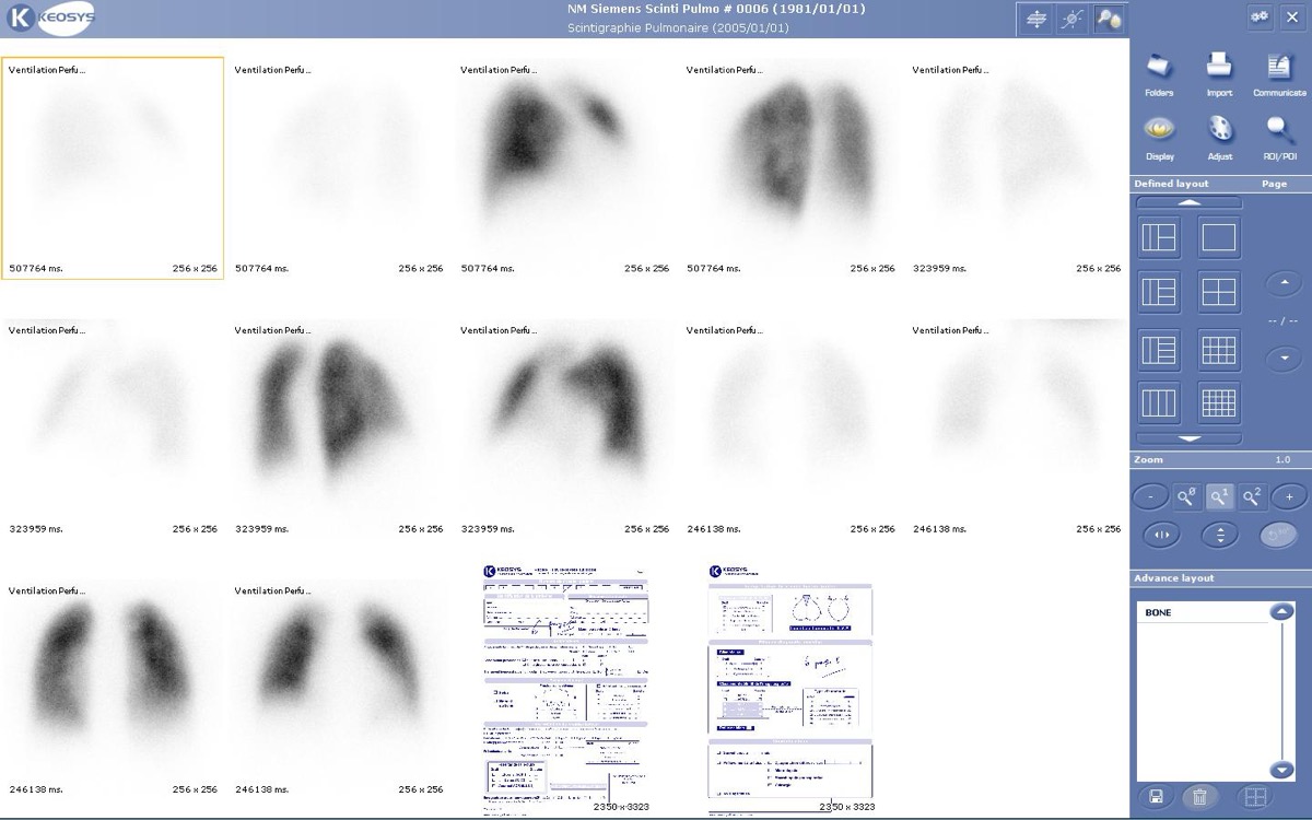 How Tc-99m MAA Lung Perfusion Imaging Works
Reducing MAA Particles in Pulmonary Hypertension
All 130 nuclear medicine images by category →
Practise image interpretation in the app
All 130 images as an interactive quiz, alongside 500 NMTCB and ARRT(N) questions with worked explanations, case studies and timed mock exams. 25 questions are free, no card and no account.