Lung Perfusion Projections: Why Lateral Views Matter
CC BY-SA 4.0 · Wikimedia Commons · source file
Question
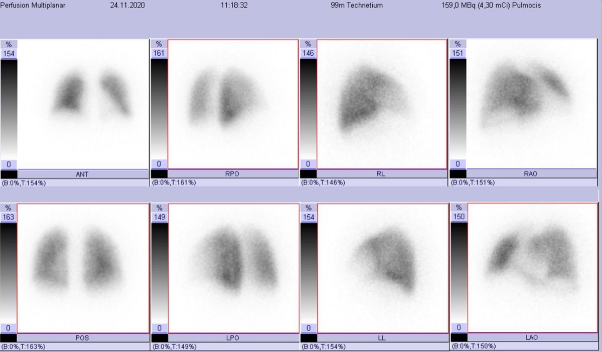
A lung perfusion scan is routinely performed in multiple projections (anterior, posterior, right and left lateral, right and left posterior oblique, right and left anterior oblique = 8 views). Why is it important to image in MULTIPLE projections rather than just anterior and posterior?
- AMultiple projections allow calculation of quantitative lung volume
- BPosterior projection alone is sufficient; multiple views are redundant
- CLateral views allow detection of posterior and anterior segment defects that overlap on AP projections, improving detection and classification of PE
- DMultiple projections allow detection of ventilation defects
Show the answer and explanation
C. Lateral views allow detection of posterior and anterior segment defects that overlap on AP projections, improving detection and classification of PE
Multiple projections (8 standard views) are essential for V/Q lung scan interpretation because lung segments that overlap on anterior/posterior views are separated on lateral and oblique views. For example, the posterior basal and lateral basal segments of the lower lobes overlap each other on the posterior view but are separated on the lateral projections. Multiple views also allow more accurate classification of defect size (segmental vs. subsegmental) and extent, which directly determines PIOPED probability category. A single-view study significantly underestimates the number of perfusion defects and can lead to incorrect classification.
More lung v/q imaging images
 High-Probability V/Q Scan: Mismatched Defects
 Matched V/Q Defects: Low Probability for PE
Normal Lung Perfusion Scan and Pulmonary Embolism
 How Tc-99m MAA Lung Perfusion Imaging Works
Reducing MAA Particles in Pulmonary Hypertension
Stripe Sign on a Lung Perfusion Scan
All 130 nuclear medicine images by category →
Practise image interpretation in the app
All 130 images as an interactive quiz, alongside 500 NMTCB and ARRT(N) questions with worked explanations, case studies and timed mock exams. 25 questions are free, no card and no account.