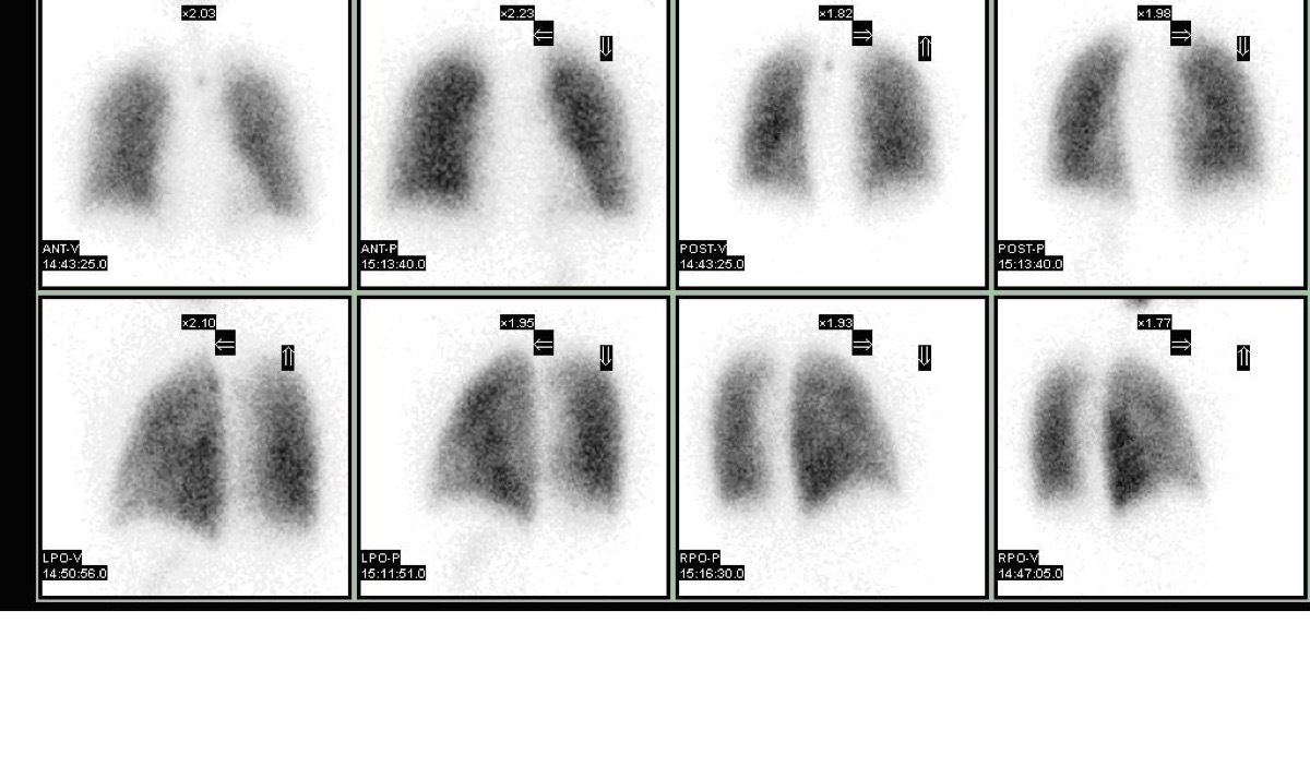
Intermediate-Probability V/Q Scan
CC BY 3.0 · Myohan / Wikimedia Commons · source file
Question
A V/Q scan performed for suspected PE shows a single moderate-sized (between 25% and 75% of a segment) ventilation-perfusion mismatch. By PIOPED II criteria, this single moderate mismatch is classified as:
- AHigh probability for PE
- BIntermediate (indeterminate) probability for PE
- CLow probability for PE
- DNormal — no PE
Show the answer and explanation
B. Intermediate (indeterminate) probability for PE
By PIOPED II criteria, INTERMEDIATE (indeterminate) probability includes: one moderate V/Q mismatch without corresponding chest X-ray abnormality; difficult-to-classify patterns not meeting high or low probability criteria; and studies with both high-probability and low-probability features. Intermediate probability scans are clinically challenging because approximately 30–40% of patients with intermediate scans have PE. Management depends on clinical pretest probability: low clinical probability + intermediate scan may allow withholding anticoagulation; high clinical probability + intermediate scan typically warrants further evaluation (CTPA) or empiric anticoagulation.
More lung v/q imaging images
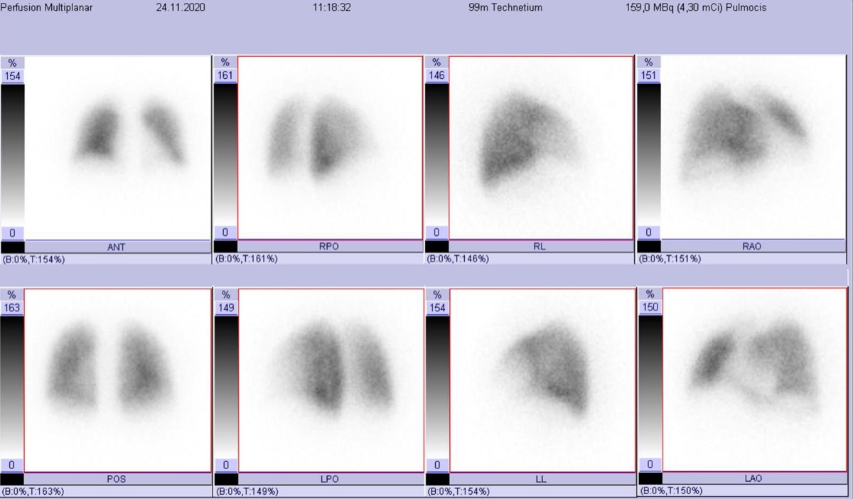 Brain and Kidney MAA Uptake: Right-to-Left Shunt
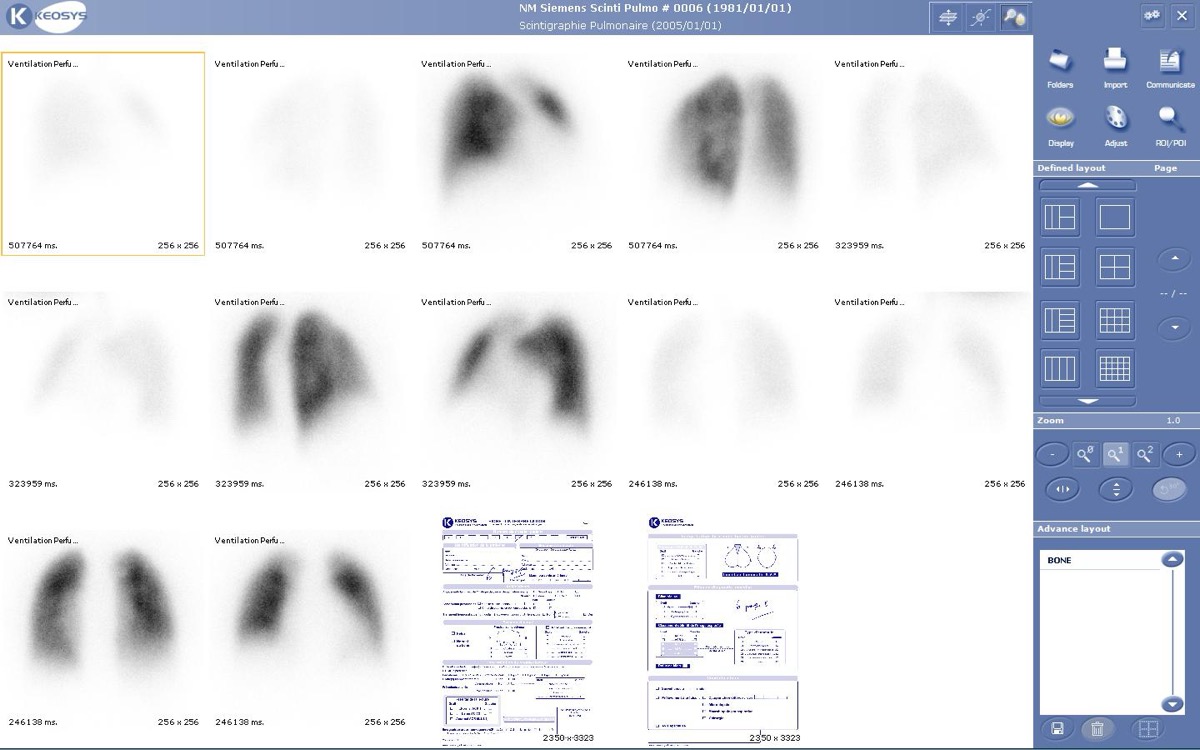 Tc-99m MAA Injection Technique
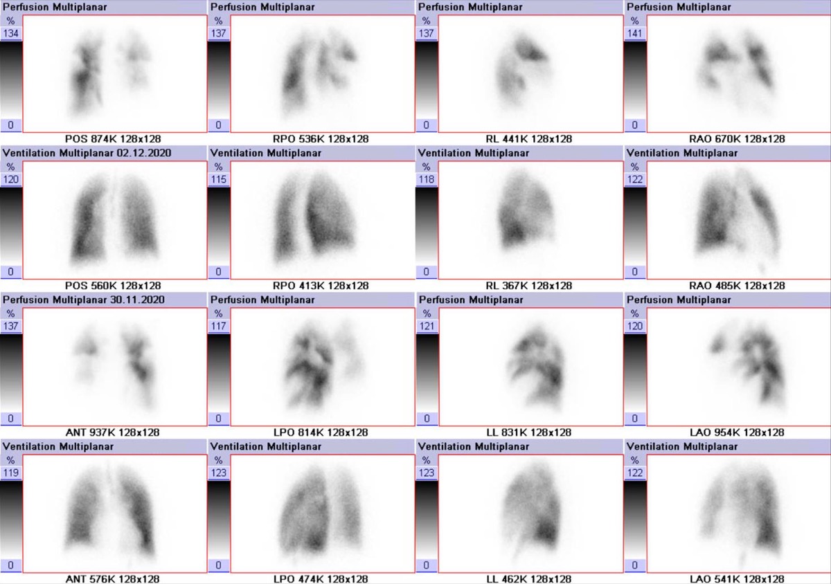 PIOPED II: High-Probability V/Q with High Pretest Probability
Wedge-Shaped Pleural-Based Defect: Pulmonary Embolism
Lung Perfusion Projections: Why Lateral Views Matter
High-Probability V/Q Scan: Mismatched Defects
All 130 nuclear medicine images by category →
Practise image interpretation in the app
All 130 images as an interactive quiz, alongside 500 NMTCB and ARRT(N) questions with worked explanations, case studies and timed mock exams. 25 questions are free, no card and no account.