Brain and Kidney MAA Uptake: Right-to-Left Shunt
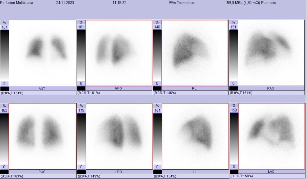
CC BY-SA 4.0 · Wikimedia Commons · source file
Question
During a V/Q scan, uptake of Tc-99m MAA is unexpectedly visualized in the brain and kidneys on the perfusion images. What does this finding indicate?
- ARight-to-left cardiac or pulmonary shunt — MAA particles are crossing from the right to the left side of the circulation
- BFree pertechnetate contamination of the MAA preparation
- CNormal thyroid and salivary gland activity from free pertechnetate
- DBilateral PE with systemic embolization
Show the answer and explanation
A. Right-to-left cardiac or pulmonary shunt — MAA particles are crossing from the right to the left side of the circulation
Tc-99m MAA particles are normally too large (10–100 μm) to pass through the pulmonary capillaries (5–10 μm), so they lodge in the lungs. If MAA activity is seen in the brain and kidneys on perfusion images, it means the particles bypassed the pulmonary capillary bed by crossing through an intracardiac shunt (patent foramen ovale, ASD, VSD) or a pulmonary arteriovenous malformation (AVM) — a right-to-left shunt. This incidental finding on a V/Q scan is diagnostically important and can prompt further evaluation with echocardiography. The degree of systemic activity can quantify the shunt fraction.
More lung v/q imaging images
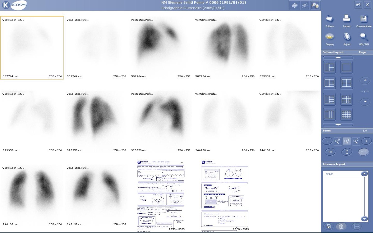 Tc-99m MAA Injection Technique
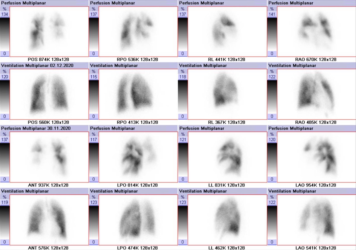 PIOPED II: High-Probability V/Q with High Pretest Probability
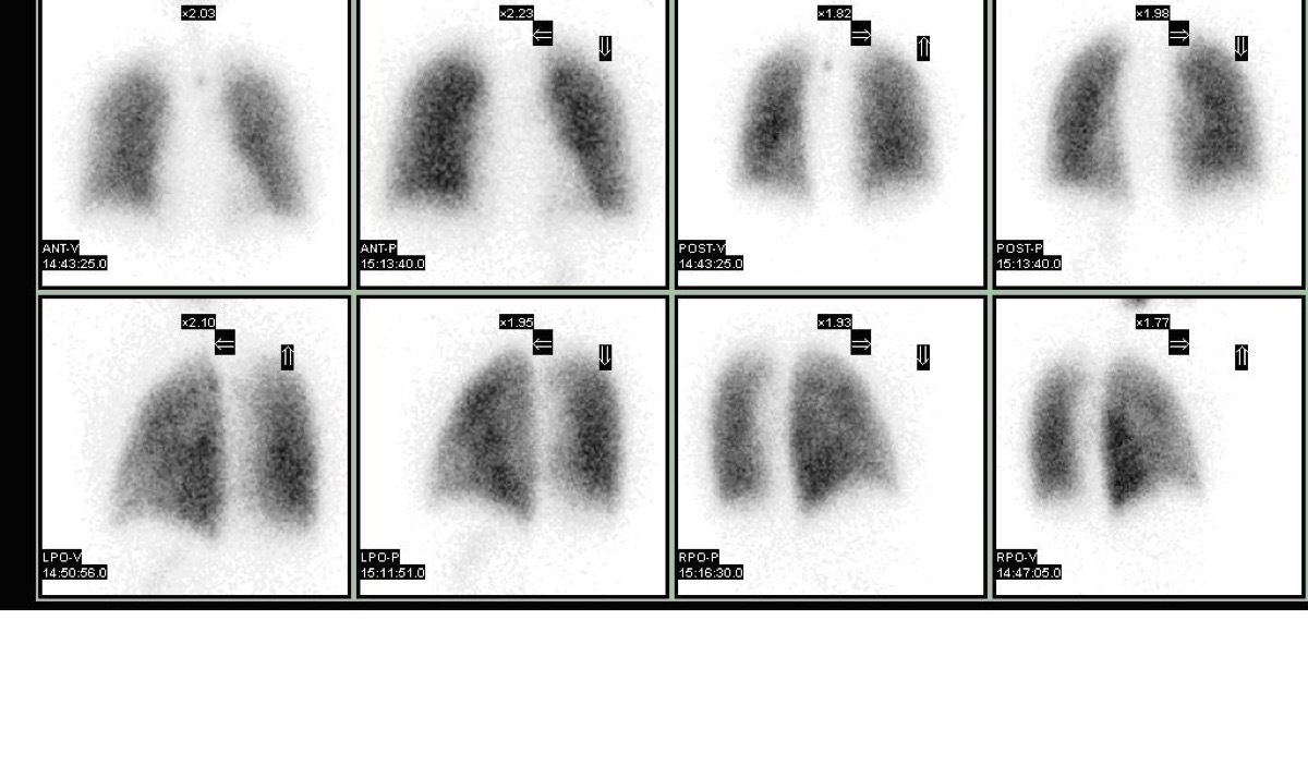 Wedge-Shaped Pleural-Based Defect: Pulmonary Embolism
Lung Perfusion Projections: Why Lateral Views Matter
High-Probability V/Q Scan: Mismatched Defects
Matched V/Q Defects: Low Probability for PE
All 130 nuclear medicine images by category →
Practise image interpretation in the app
All 130 images as an interactive quiz, alongside 500 NMTCB and ARRT(N) questions with worked explanations, case studies and timed mock exams. 25 questions are free, no card and no account.