PIOPED II: High-Probability V/Q with High Pretest Probability
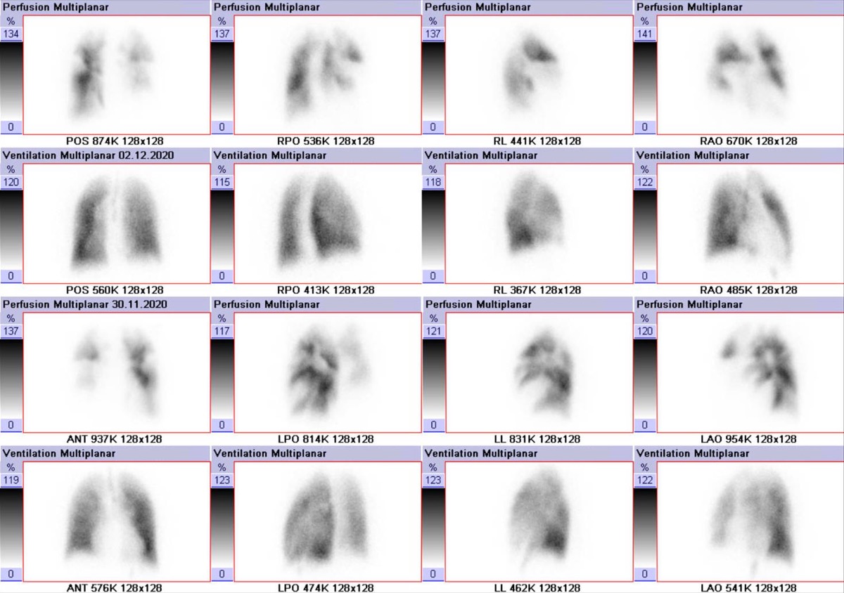
CC BY-SA 4.0 · Hellerhoff / Wikimedia Commons · source file
Question
According to PIOPED II criteria, which V/Q scan result, combined with HIGH clinical pre-test probability, effectively CONFIRMS pulmonary embolism without need for additional imaging?
- AIntermediate probability V/Q scan
- BHigh probability V/Q scan — combined with high pre-test probability, the positive predictive value for PE approaches 96%
- CLow probability V/Q scan
- DNormal V/Q scan
Show the answer and explanation
B. High probability V/Q scan — combined with high pre-test probability, the positive predictive value for PE approaches 96%
When a HIGH probability V/Q scan is combined with HIGH clinical pre-test probability (e.g., Wells score high risk), the positive predictive value (PPV) for PE approaches 96%. This combination is considered sufficient to diagnose PE and begin anticoagulation without CTPA in most guidelines. Conversely, a NORMAL V/Q scan combined with LOW pre-test probability effectively rules out PE (NPV ~99%). The V/Q scan's clinical utility depends on integrating scan result with pre-test probability: intermediate scans in intermediate-probability patients represent the most diagnostically challenging scenario and often require CTPA.
More lung v/q imaging images
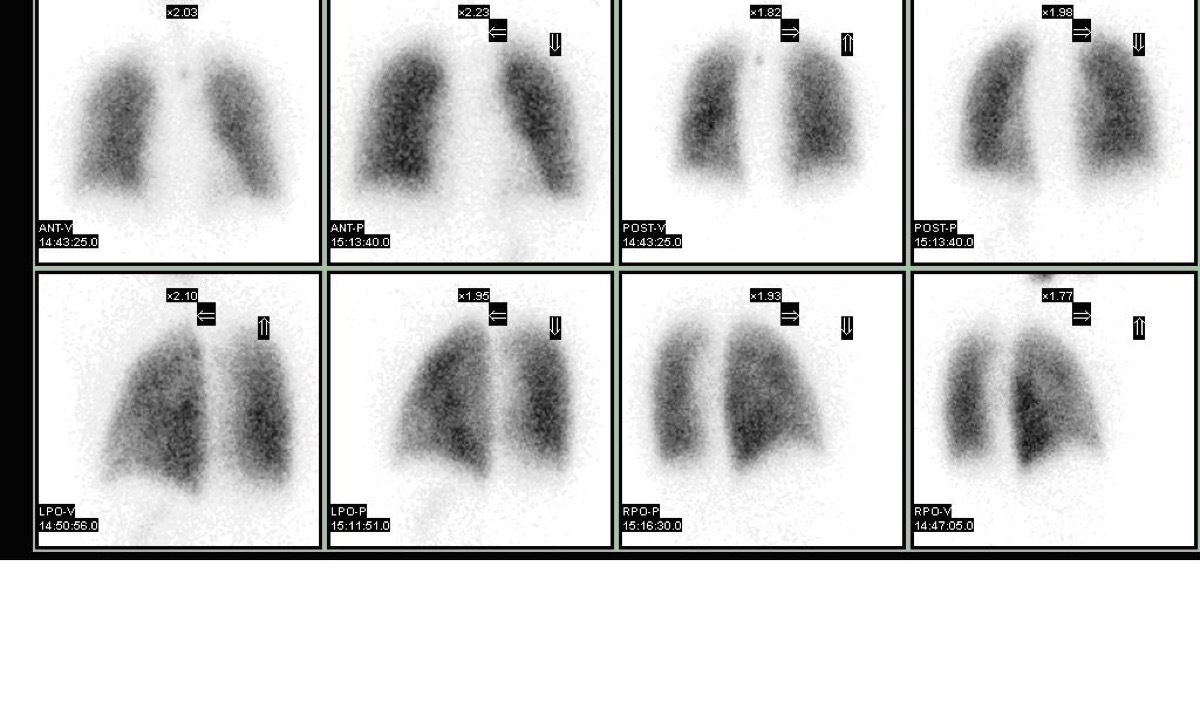 Wedge-Shaped Pleural-Based Defect: Pulmonary Embolism
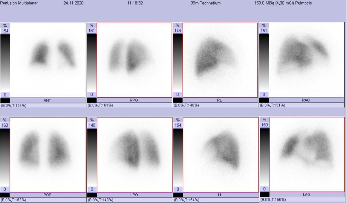 Lung Perfusion Projections: Why Lateral Views Matter
High-Probability V/Q Scan: Mismatched Defects
Matched V/Q Defects: Low Probability for PE
Normal Lung Perfusion Scan and Pulmonary Embolism
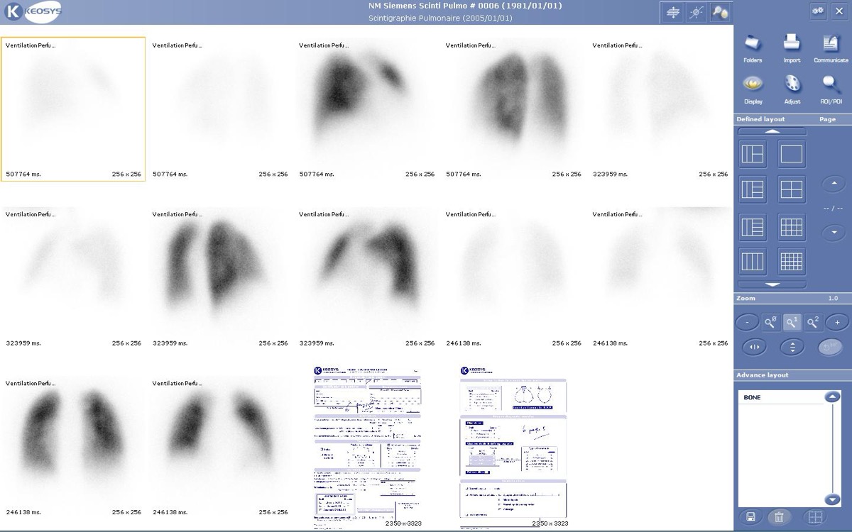 How Tc-99m MAA Lung Perfusion Imaging Works
All 130 nuclear medicine images by category →
Practise image interpretation in the app
All 130 images as an interactive quiz, alongside 500 NMTCB and ARRT(N) questions with worked explanations, case studies and timed mock exams. 25 questions are free, no card and no account.