V/Q Scan in COPD: Matched Defects
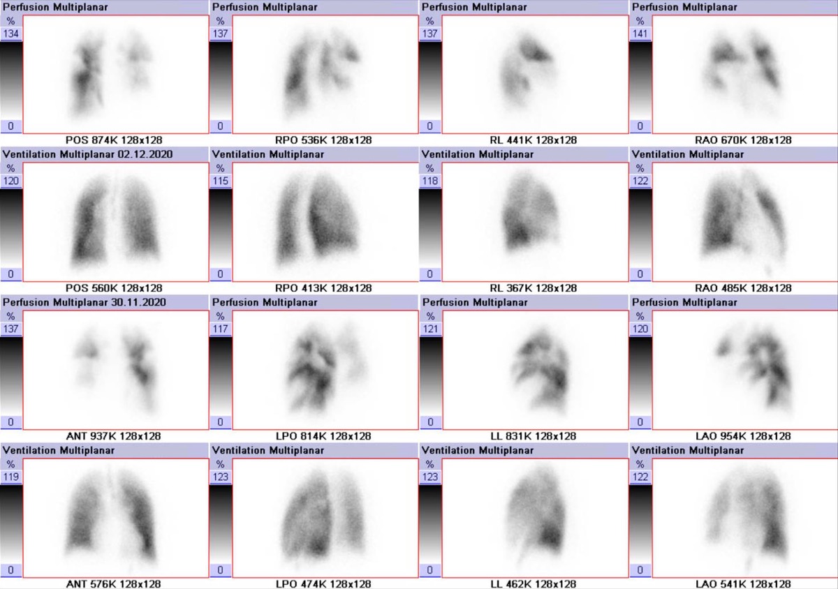
CC BY-SA 4.0 · Hellerhoff / Wikimedia Commons · source file
Question
A patient with severe COPD undergoes V/Q scanning to evaluate for PE. The scan shows multiple matched ventilation and perfusion defects throughout both lung fields. By PIOPED II criteria, this pattern is classified as:
- ALow probability for PE — matched defects in COPD are characteristic of parenchymal disease, not vascular obstruction
- BHigh probability for PE
- CNormal study
- DCertain diagnosis of PE
Show the answer and explanation
A. Low probability for PE — matched defects in COPD are characteristic of parenchymal disease, not vascular obstruction
In patients with COPD, areas of airway obstruction reduce both ventilation AND perfusion to the same lung regions (hypoxic vasoconstriction and/or loss of vascular bed), producing matched V/Q defects. By PIOPED II criteria, matched defects — particularly when they correspond to abnormalities on chest X-ray — represent LOW probability for PE. The V/Q scan is less specific in patients with COPD because the underlying lung disease creates matched defects that mimic the appearance of PE-related mismatches but without actual vascular occlusion. CT pulmonary angiography (CTPA) is often preferred in patients with severe COPD.
More lung v/q imaging images
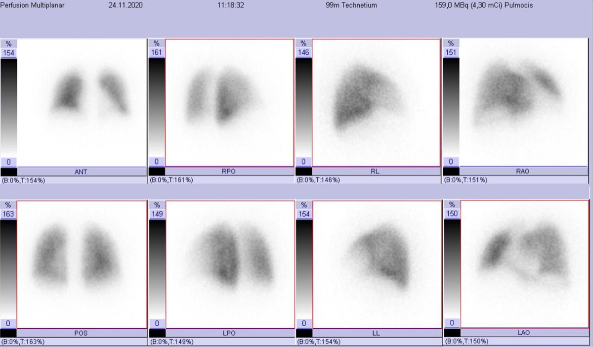 V/Q Scan vs CTPA for Pulmonary Embolism in Pregnancy
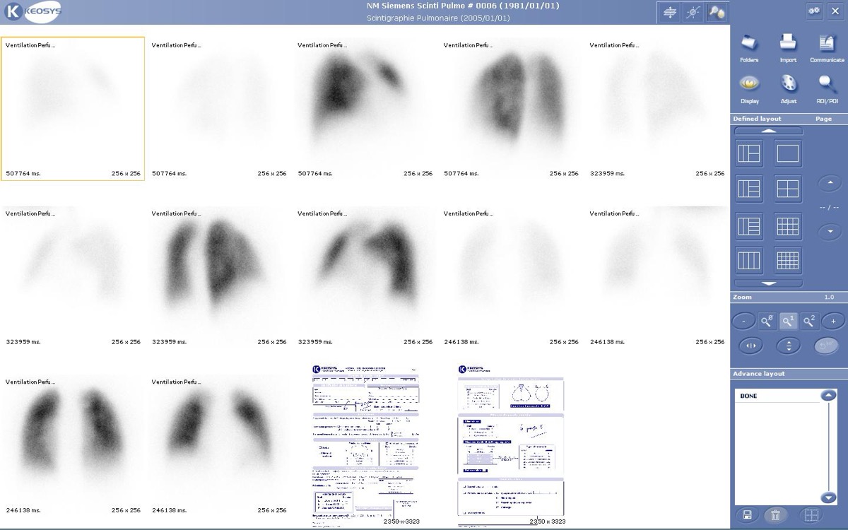 Ventilation Agents: Tc-99m DTPA Aerosol vs Xe-133
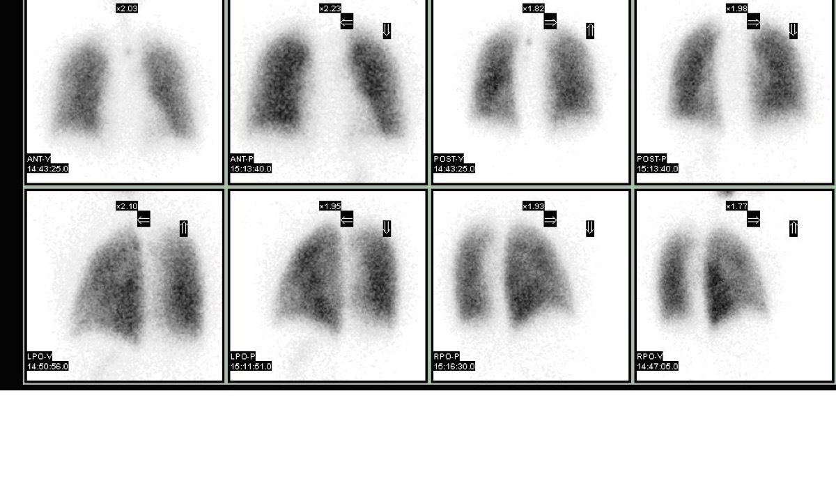 Intermediate-Probability V/Q Scan
Brain and Kidney MAA Uptake: Right-to-Left Shunt
Tc-99m MAA Injection Technique
PIOPED II: High-Probability V/Q with High Pretest Probability
All 130 nuclear medicine images by category →
Practise image interpretation in the app
All 130 images as an interactive quiz, alongside 500 NMTCB and ARRT(N) questions with worked explanations, case studies and timed mock exams. 25 questions are free, no card and no account.