Stripe Sign on a Lung Perfusion Scan
CC BY 3.0 · Myohan / Wikimedia Commons · source file
Question
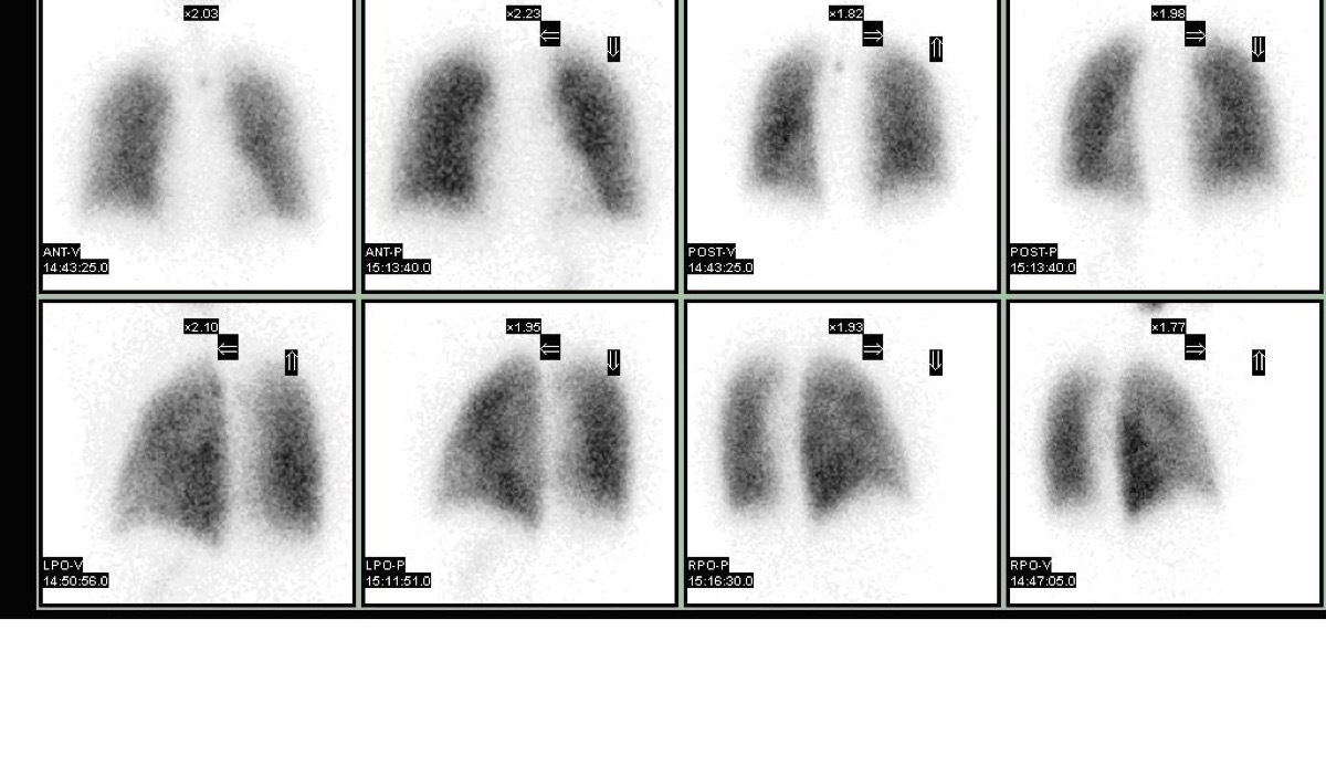
On a lung perfusion scan, the 'stripe sign' is a finding where a thin rim of perfusion is preserved along the pleural edge of a peripheral lung zone that appears otherwise hypo-perfused. The stripe sign is important because:
- AIt indicates a high probability pattern for PE
- BIt confirms bilateral PE
- CIt represents a technical artifact from motion
- DIt indicates the defect is likely NOT due to PE — the preserved peripheral perfusion stripe argues against vascular occlusion
Show the answer and explanation
D. It indicates the defect is likely NOT due to PE — the preserved peripheral perfusion stripe argues against vascular occlusion
The 'stripe sign' refers to a thin rim of preserved perfusion at the pleural surface adjacent to a perfusion defect. Since pulmonary emboli occlude vessels that supply the lung parenchyma (including the subpleural region), a true PE-related defect would typically extend to the pleural edge without a preserved peripheral stripe. The presence of a perfusion stripe suggests that the defect is caused by parenchymal disease (atelectasis, mucus plug, COPD) that affects the central lung but spares the most peripheral subpleural perfusion via collateral flow. The stripe sign is associated with a lower probability interpretation per PIOPED criteria.
More lung v/q imaging images
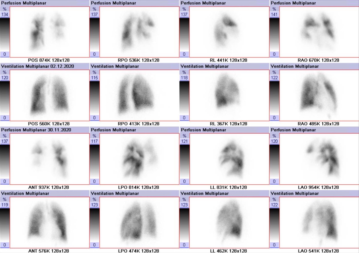 V/Q Scan in COPD: Matched Defects
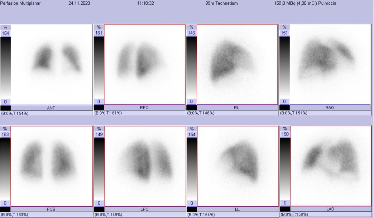 V/Q Scan vs CTPA for Pulmonary Embolism in Pregnancy
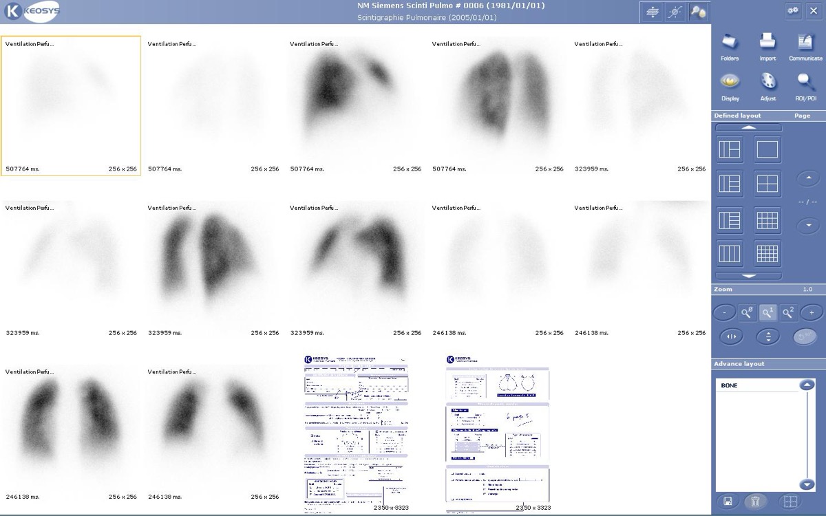 Ventilation Agents: Tc-99m DTPA Aerosol vs Xe-133
Intermediate-Probability V/Q Scan
Brain and Kidney MAA Uptake: Right-to-Left Shunt
Tc-99m MAA Injection Technique
All 130 nuclear medicine images by category →
Practise image interpretation in the app
All 130 images as an interactive quiz, alongside 500 NMTCB and ARRT(N) questions with worked explanations, case studies and timed mock exams. 25 questions are free, no card and no account.