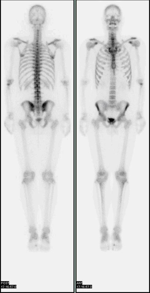
Shin Splints on Bone Scan: Medial Tibial Stress Syndrome
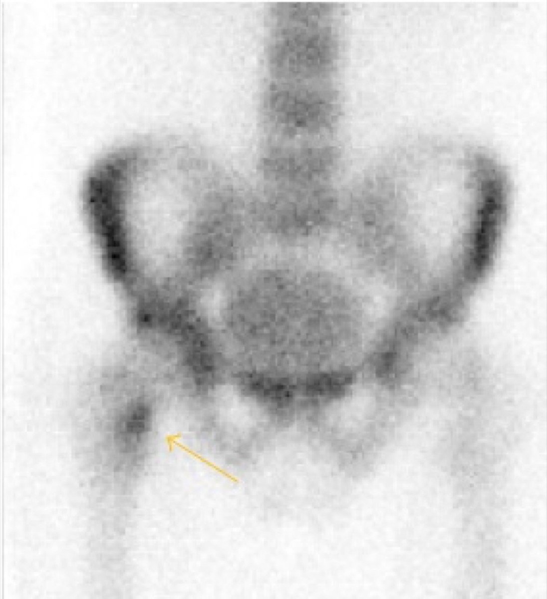
CC BY 4.0 · Santiago et al. / Wikimedia Commons · source file
Question
A bone scan of a distance runner with medial lower leg pain shows a linear band of mildly increased uptake along the posteromedial cortex of the tibia, extending over a long segment. This pattern is most consistent with:
- AMedial tibial stress syndrome (shin splints)
- BComplete tibial stress fracture
- CTibial osteosarcoma
- DOsteomyelitis of the tibia
Show the answer and explanation
A. Medial tibial stress syndrome (shin splints)
Medial tibial stress syndrome (MTSS, 'shin splints') produces a characteristic longitudinal band of mildly increased uptake along the posteromedial cortex of the tibia on bone scan, representing periosteal stress reaction over a long segment. This distinguishes it from a stress fracture, which shows a more focal, fusiform, transverse focus of intense uptake at the cortex. MTSS uptake is also typically less intense than a true stress fracture. The three-phase bone scan shows no significant flow or blood pool abnormality in MTSS (unlike stress fracture or osteomyelitis).
More bone imaging images
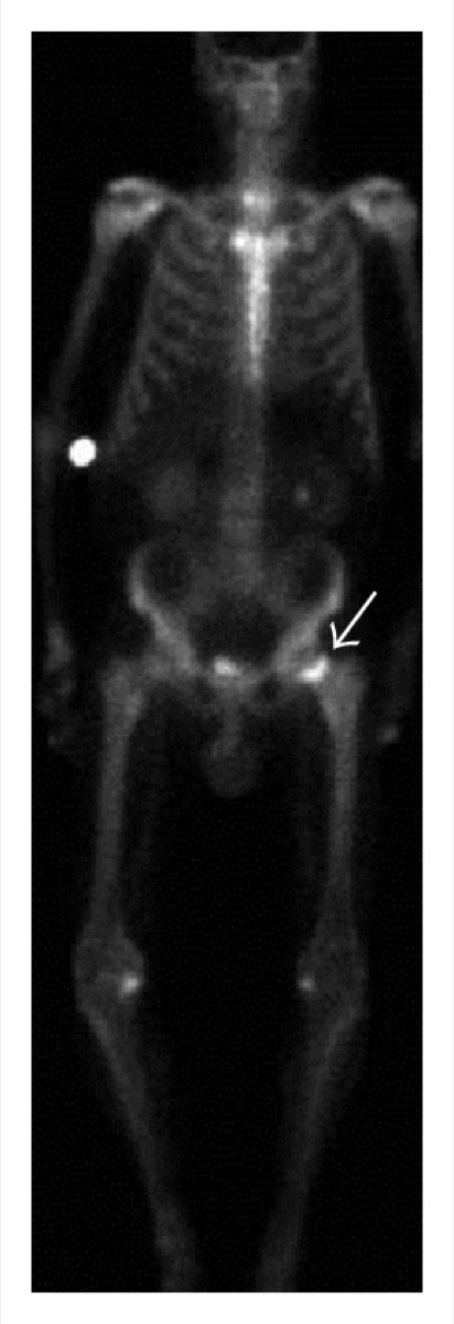 Avascular Necrosis of the Femoral Head on Bone Scan
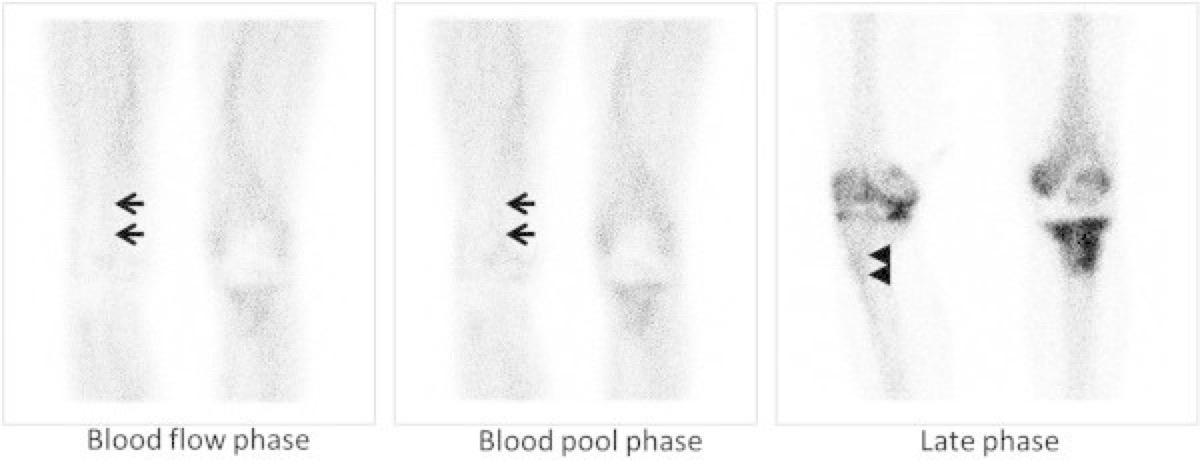 Flare Phenomenon on Bone Scan After Chemotherapy
 Bone Scan for Staging High-Risk Prostate Cancer
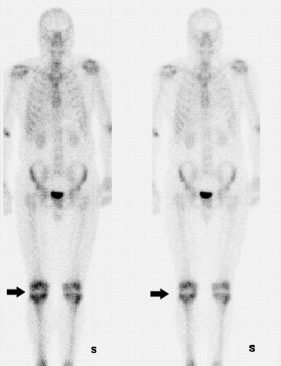 Fibrous Dysplasia on Bone Scan (Shepherd’s Crook)
 Tc-99m Physical Half-Life
Periprosthetic Uptake After Hip Replacement
All 130 nuclear medicine images by category →
Practise image interpretation in the app
All 130 images as an interactive quiz, alongside 500 NMTCB and ARRT(N) questions with worked explanations, case studies and timed mock exams. 25 questions are free, no card and no account.