Periprosthetic Uptake After Hip Replacement
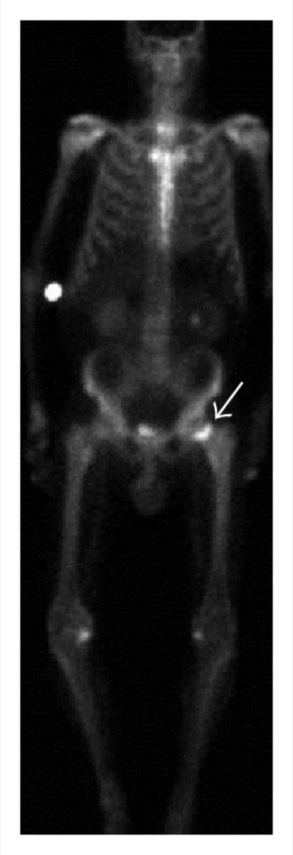
CC BY 3.0 · Jarraya et al. / Wikimedia Commons · source file
Question
A bone scan performed 1 year after total hip replacement shows mildly increased uptake around the acetabular component and femoral stem tip. At what time AFTER hip replacement does normal post-operative bone remodeling uptake typically return to baseline on bone scan?
- A6–12 months for the acetabulum and prosthetic tip — uptake beyond this suggests loosening or infection
- B1 week post-operatively
- CUptake never resolves and is always present around prostheses
- D24–48 hours
Show the answer and explanation
A. 6–12 months for the acetabulum and prosthetic tip — uptake beyond this suggests loosening or infection
After total hip replacement (THR), normal post-operative bone remodeling causes increased uptake around the prosthesis for up to 6–12 months (sometimes 12–24 months for cemented prostheses). Uptake that PERSISTS beyond this expected time frame — particularly at the acetabulum, calcar, or femoral stem tip — raises concern for aseptic loosening, infection (periprosthetic joint infection), or stress fracture. Three-phase bone scan combined with labeled white blood cell (WBC) scan or In-111 WBC scan helps distinguish aseptic loosening from infection.
More bone imaging images
 Bone Scan Timing: When to Acquire Delayed Images
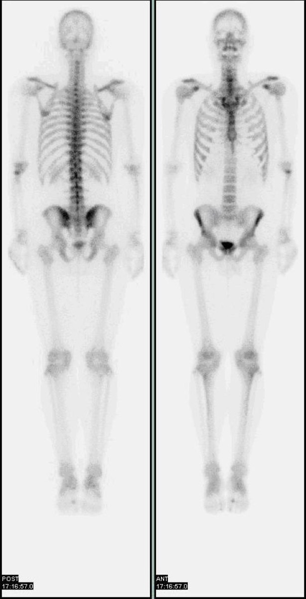 Normal Whole-Body Bone Scan (Tc-99m MDP)
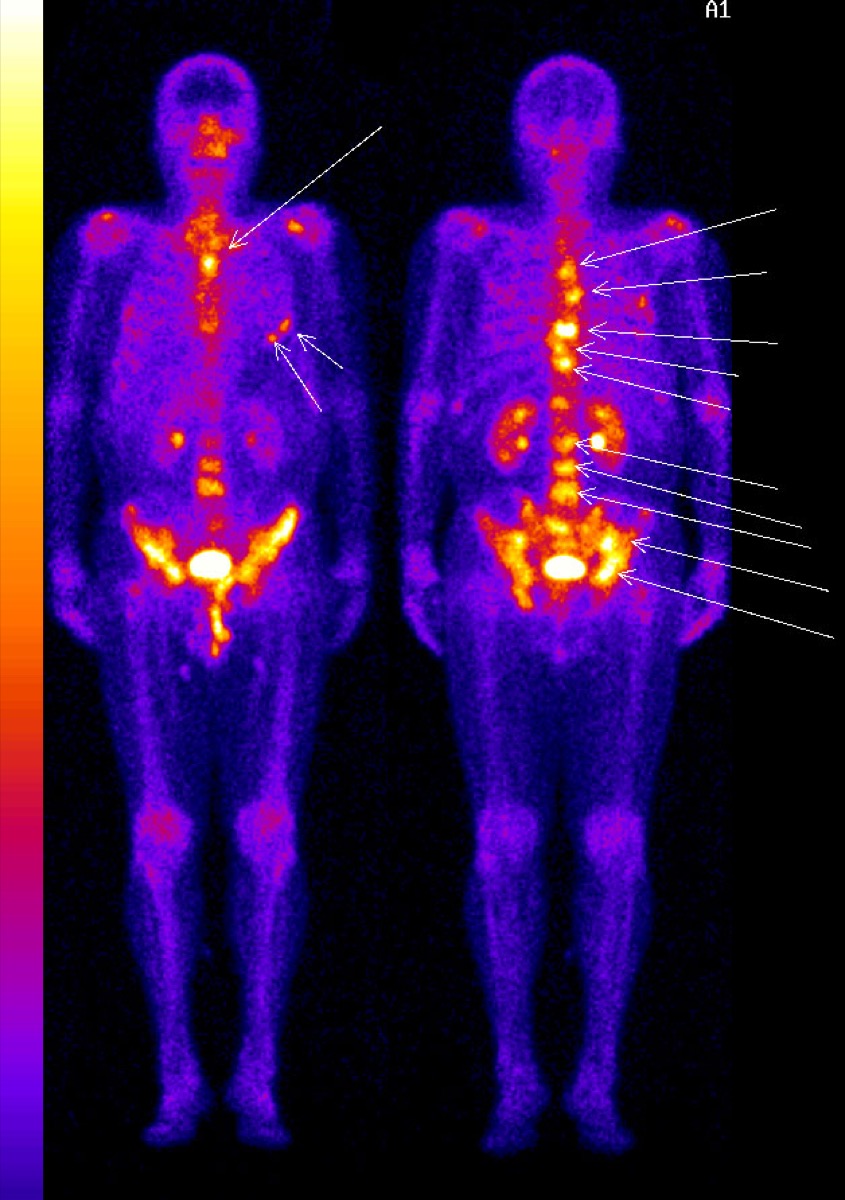 Diffuse Skeletal Metastases on Bone Scan
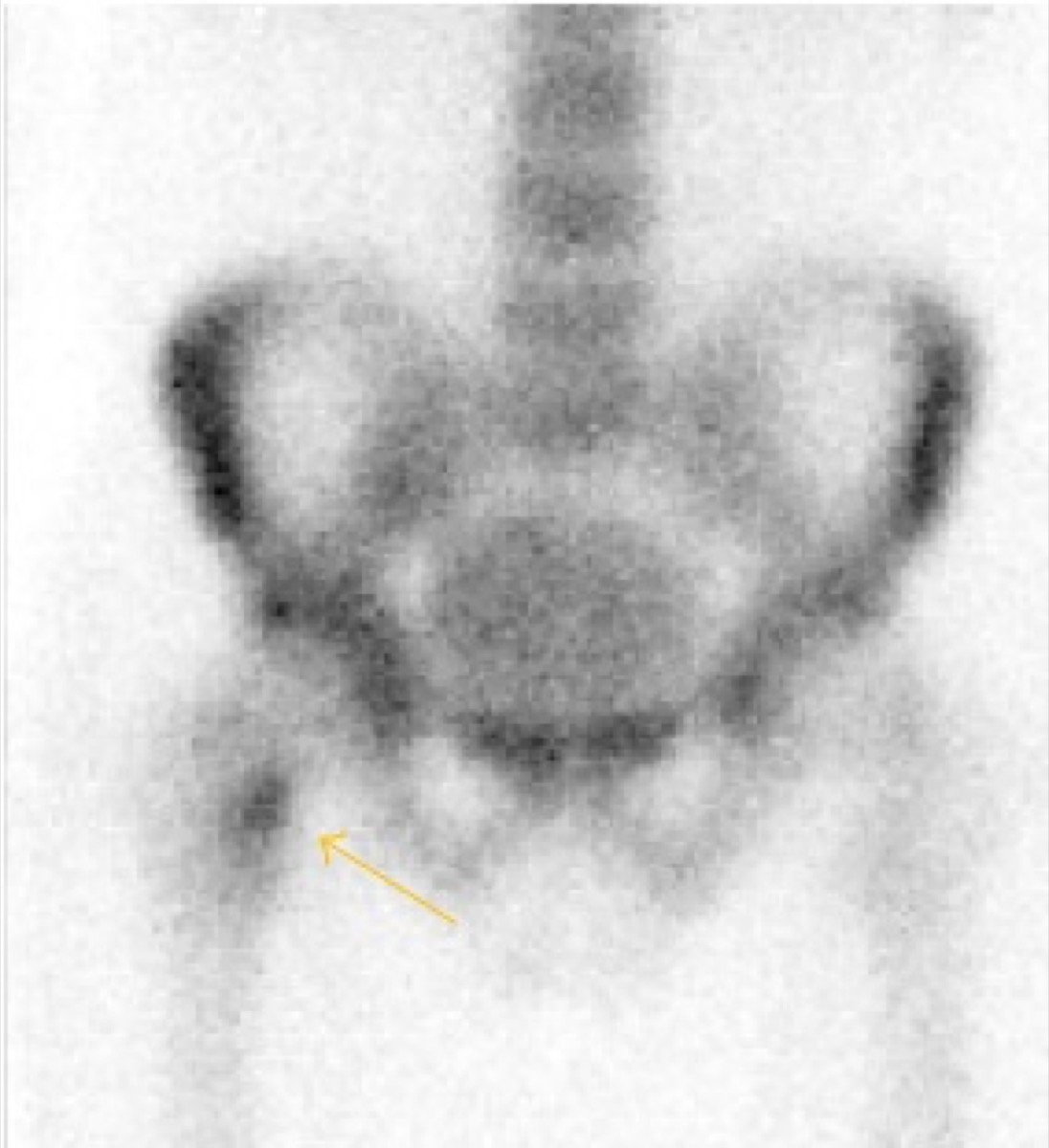 Femoral Neck Stress Fracture on Bone Scan
Femoral Neck Insufficiency Fracture on Bone Scan
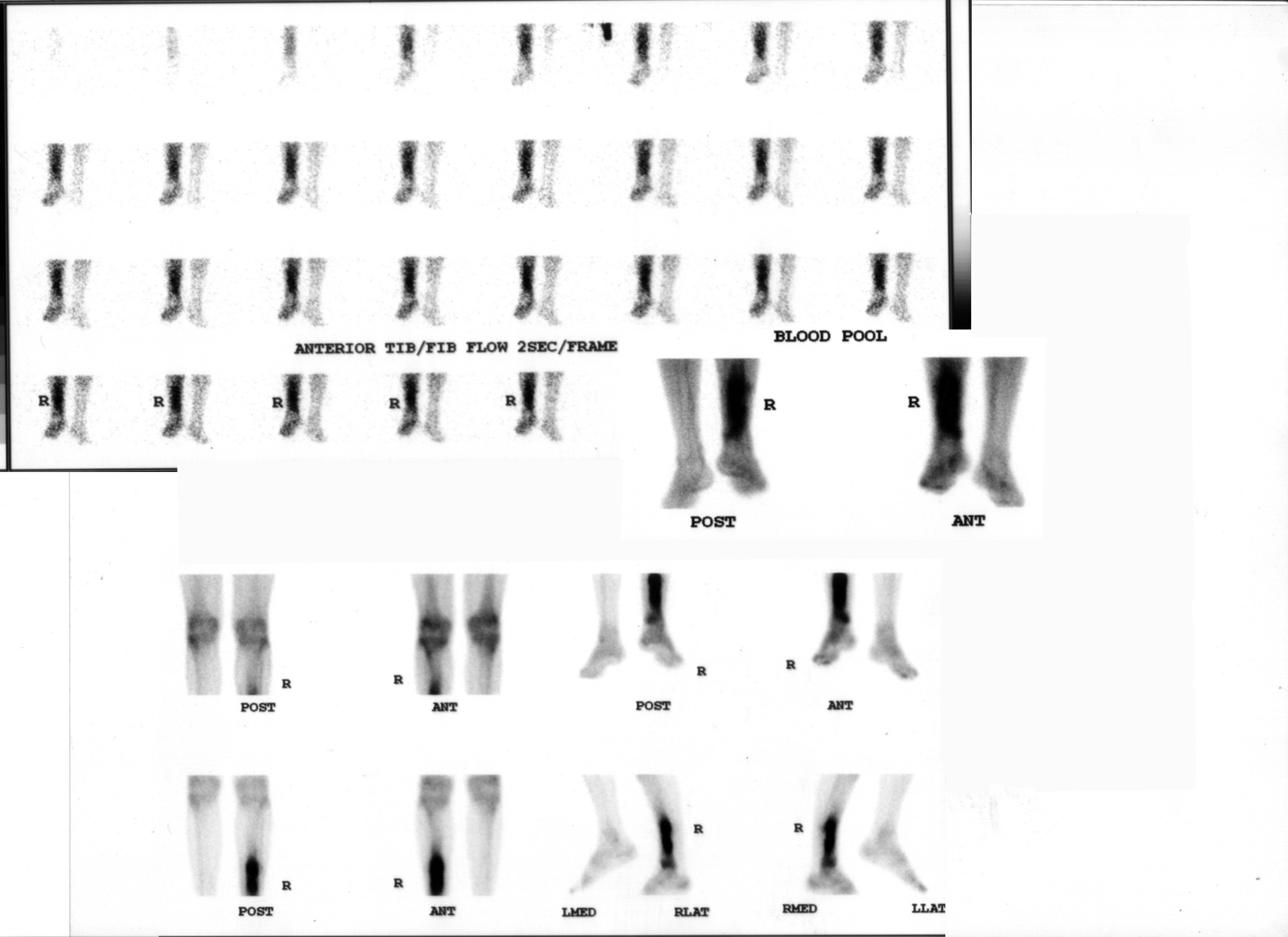 Osteomyelitis on Three-Phase Bone Scan
All 130 nuclear medicine images by category →
Practise image interpretation in the app
All 130 images as an interactive quiz, alongside 500 NMTCB and ARRT(N) questions with worked explanations, case studies and timed mock exams. 25 questions are free, no card and no account.