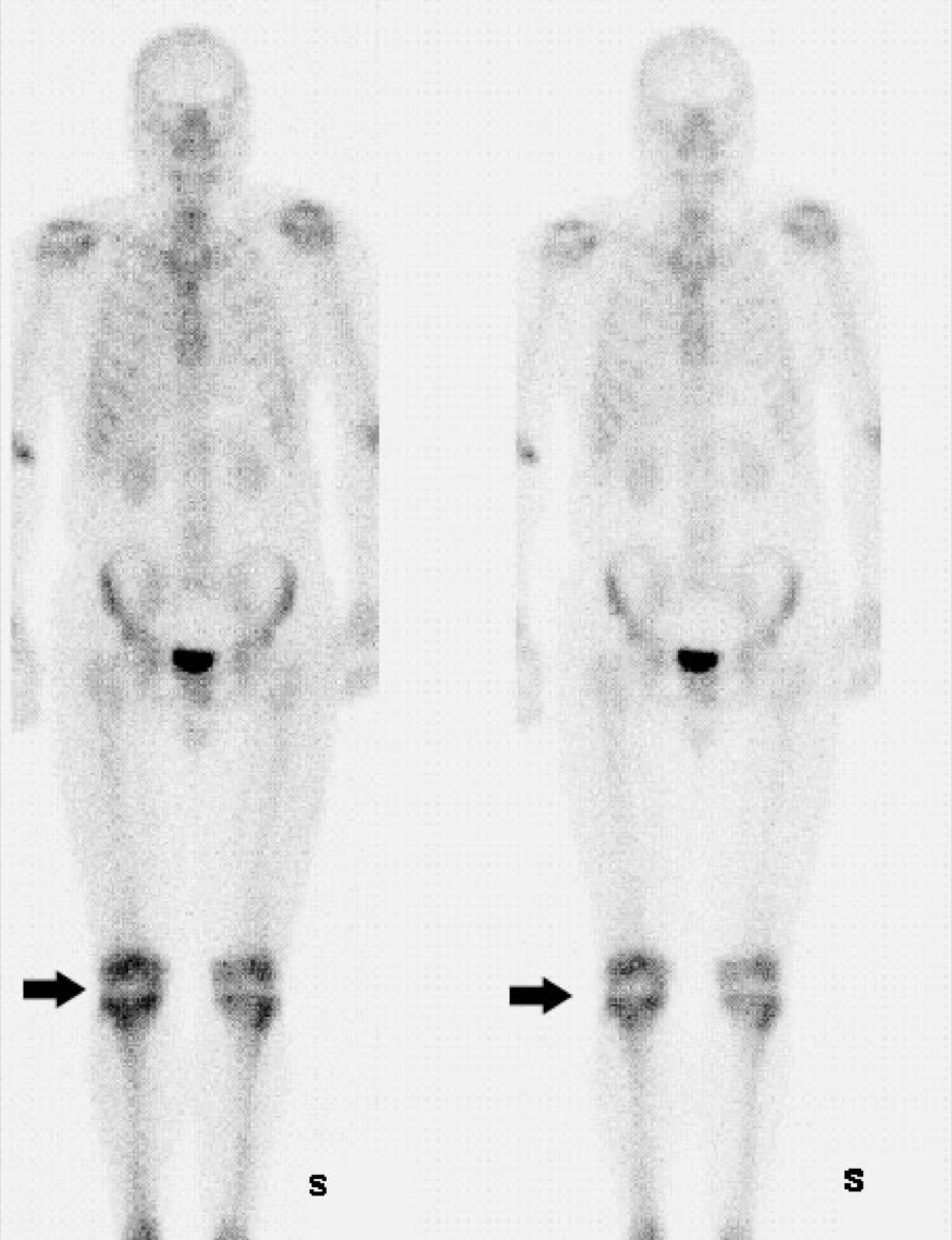
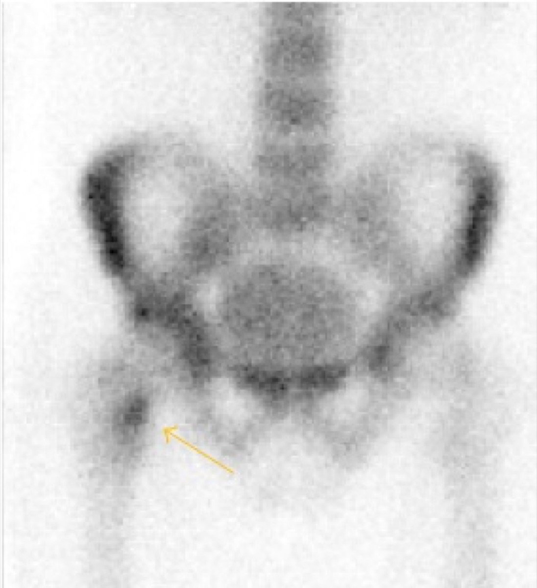
Fibrous Dysplasia on Bone Scan (Shepherd’s Crook)
CC BY-SA 3.0 · Wikimedia Commons · source file
Question
A bone scan in a young patient shows intense focal uptake in the proximal femoral shaft with a corresponding curved ('shepherd's crook') deformity on radiograph. No malignancy is suspected. This pattern is most consistent with:
- AOsteosarcoma of the femur
- BEwing's sarcoma
- CStress fracture with valgus deformity
- DFibrous dysplasia — monostotic or polyostotic form
Show the answer and explanation
D. Fibrous dysplasia — monostotic or polyostotic form
Fibrous dysplasia is a benign skeletal disorder in which normal bone is replaced by fibrous tissue with immature woven bone. It commonly affects the proximal femur, producing the classic 'shepherd's crook' deformity (coxa vara). On bone scan, fibrous dysplasia shows moderate-to-intense increased uptake proportional to the degree of bone turnover and lesion activity. Bone scan is useful for determining the full extent of disease (monostotic vs. polyostotic). Unlike malignant lesions, fibrous dysplasia is a benign incidental finding in young patients, and the radiographic appearance is characteristic.
More bone imaging images
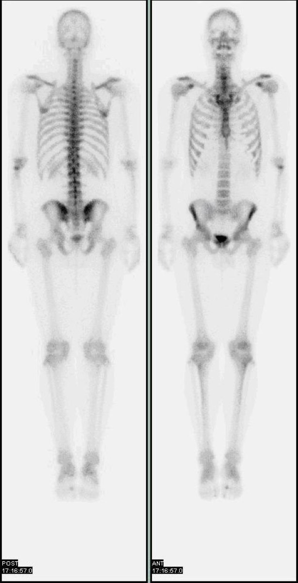 Tc-99m Physical Half-Life
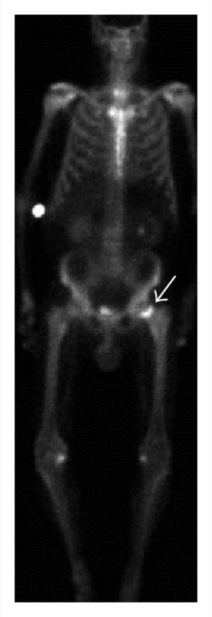 Periprosthetic Uptake After Hip Replacement
 Bone Scan Timing: When to Acquire Delayed Images
Normal Whole-Body Bone Scan (Tc-99m MDP)
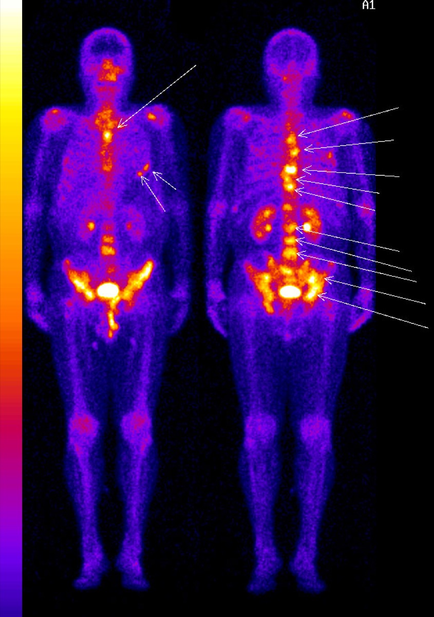 Diffuse Skeletal Metastases on Bone Scan
 Femoral Neck Stress Fracture on Bone Scan
All 130 nuclear medicine images by category →
Practise image interpretation in the app
All 130 images as an interactive quiz, alongside 500 NMTCB and ARRT(N) questions with worked explanations, case studies and timed mock exams. 25 questions are free, no card and no account.