Flare Phenomenon on Bone Scan After Chemotherapy
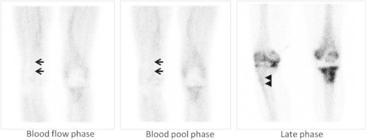
CC BY 2.0 · Wikimedia Commons · source file
Question
A patient with known breast cancer recently completed a new chemotherapy regimen. A follow-up bone scan shows MORE lesions and more intense uptake than the scan performed 3 months ago, yet tumor markers are declining. This increase in bone scan abnormalities after therapy is BEST explained by:
- AProgressive bone metastases with paradoxical tumor marker suppression due to cytokine-mediated feedback inhibition of CA 15-3 synthesis
- BDevelopment of a superscan pattern secondary to diffuse marrow infiltration with compensatory extramedullary hematopoiesis masking regional differences
- CFlare phenomenon — transient increase in bone scan activity reflecting healing osteoblastic response to effective therapy
- DLaboratory artifact from chemotherapy-induced heterophilic antibody interference with immunoassay-based tumor marker quantification
Show the answer and explanation
C. Flare phenomenon — transient increase in bone scan activity reflecting healing osteoblastic response to effective therapy
The 'flare phenomenon' is a well-recognized response to effective cancer therapy on bone scintigraphy. When chemotherapy or hormonal therapy kills tumor cells, it triggers an osteoblastic healing response in the lesion sites, causing transiently increased MDP uptake on bone scan within the first 3–6 months of treatment. This appears as new or more intense lesions despite clinical improvement and declining tumor markers. The flare phenomenon must be distinguished from true progression; a repeat scan 3–6 months later will show improvement if therapy is working.
More bone imaging images
 Bone Scan for Staging High-Risk Prostate Cancer
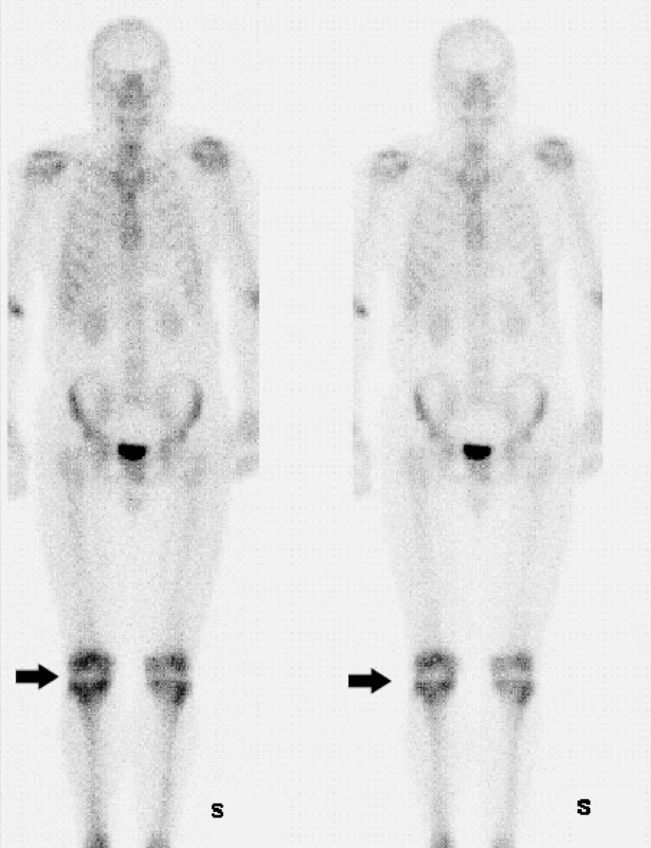 Fibrous Dysplasia on Bone Scan (Shepherd’s Crook)
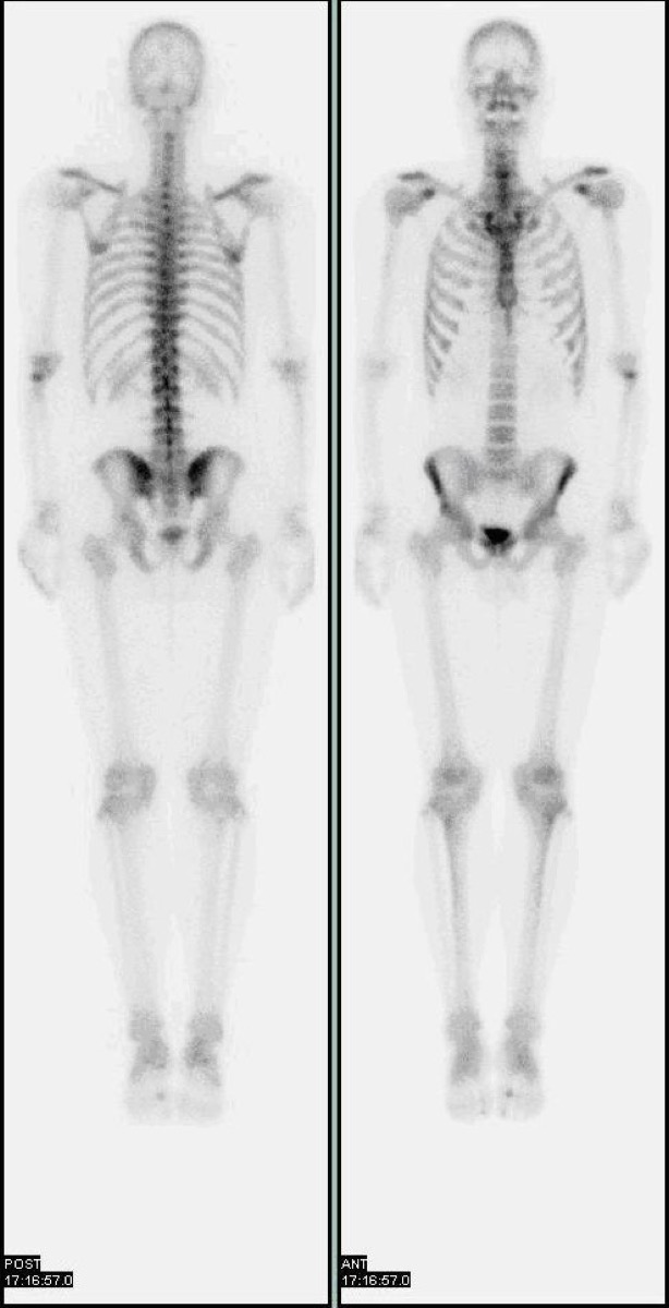 Tc-99m Physical Half-Life
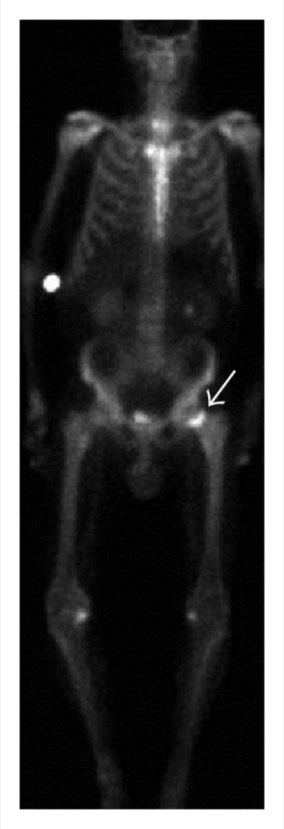 Periprosthetic Uptake After Hip Replacement
Bone Scan Timing: When to Acquire Delayed Images
Normal Whole-Body Bone Scan (Tc-99m MDP)
All 130 nuclear medicine images by category →
Practise image interpretation in the app
All 130 images as an interactive quiz, alongside 500 NMTCB and ARRT(N) questions with worked explanations, case studies and timed mock exams. 25 questions are free, no card and no account.