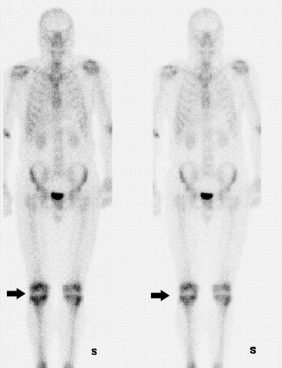
Normal Physiologic Uptake on a Bone Scan
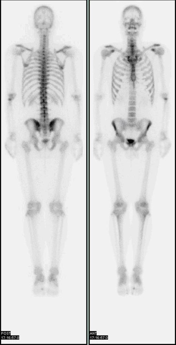
CC BY 3.0 · Myohan / Wikimedia Commons · source file
Question
On a normal Tc-99m MDP whole-body bone scan, which of the following structures normally shows PHYSIOLOGIC radiotracer activity on the delayed (bone-phase) images?
- ASpleen and thyroid
- BKidneys, bladder, and sites of active bone growth (growth plates in children)
- CBrain and liver
- DLungs and stomach
Show the answer and explanation
B. Kidneys, bladder, and sites of active bone growth (growth plates in children)
On normal delayed bone scan images, physiologic uptake is expected in: (1) KIDNEYS — MDP is excreted by glomerular filtration, so renal activity is always visible; (2) BLADDER — urine concentrates the tracer in the bladder; (3) GROWTH PLATES — in children and adolescents, actively growing epiphyses show intense physiologic uptake at all metaphyseal regions; (4) AXIAL SKELETON — moderately increased uptake in spine, sternum, ribs, and pelvis. Soft tissue activity (outside of kidney/bladder) is abnormal and suggests poor renal clearance, free pertechnetate injection, or lymphedema.
More bone imaging images
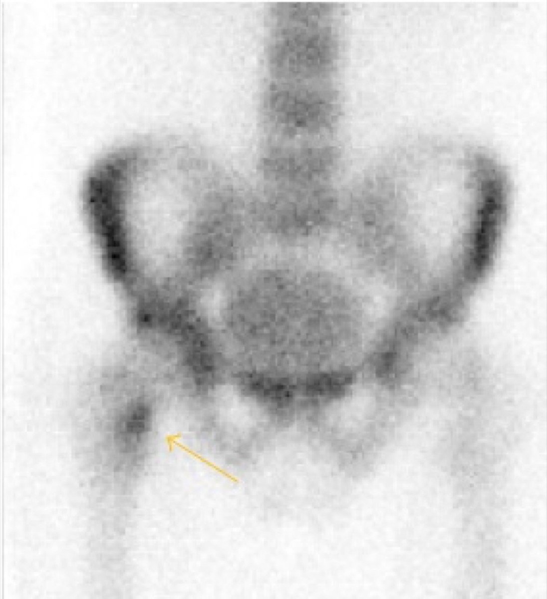 Shin Splints on Bone Scan: Medial Tibial Stress Syndrome
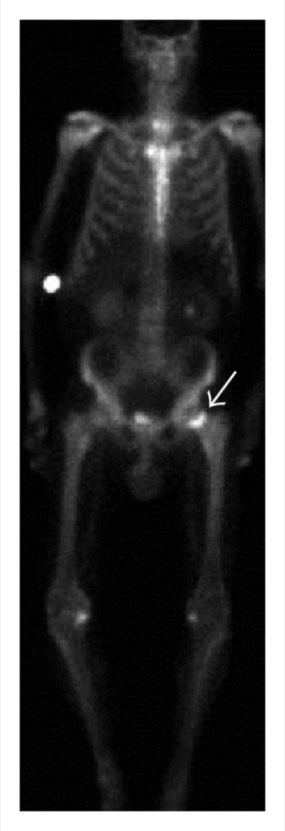 Avascular Necrosis of the Femoral Head on Bone Scan
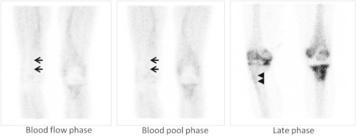 Flare Phenomenon on Bone Scan After Chemotherapy
 Bone Scan for Staging High-Risk Prostate Cancer
 Fibrous Dysplasia on Bone Scan (Shepherd’s Crook)
Tc-99m Physical Half-Life
All 130 nuclear medicine images by category →
Practise image interpretation in the app
All 130 images as an interactive quiz, alongside 500 NMTCB and ARRT(N) questions with worked explanations, case studies and timed mock exams. 25 questions are free, no card and no account.