High-Probability V/Q Scan: Mismatched Defects
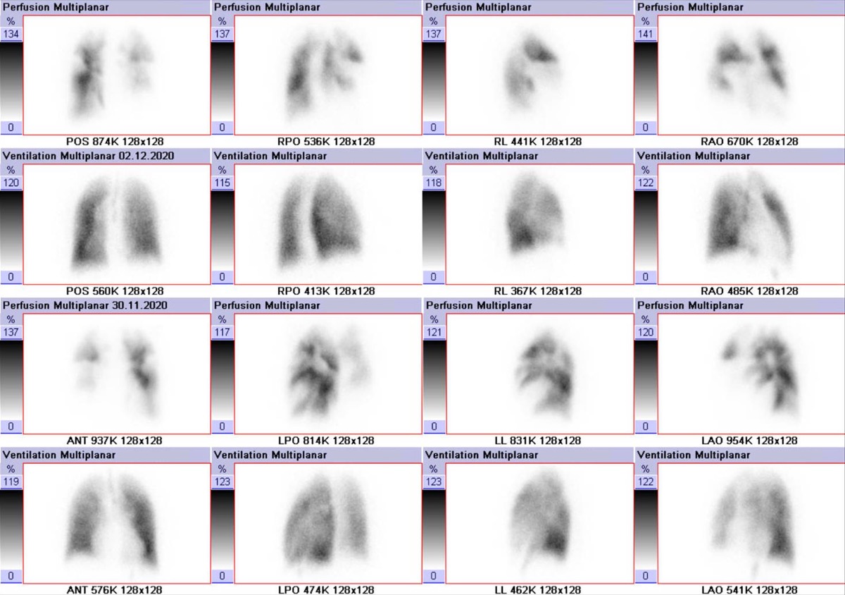
CC BY-SA 4.0 · Hellerhoff / Wikimedia Commons · source file
Question
This V/Q scan shows normal ventilation with multiple segmental/lobar perfusion defects (a 'mismatch' pattern). According to the PIOPED II criteria, this pattern is interpreted as:
- ALow probability for pulmonary embolism
- BIntermediate (indeterminate) probability for pulmonary embolism
- CHigh probability for pulmonary embolism
- DNormal — no evidence of pulmonary embolism
Show the answer and explanation
C. High probability for pulmonary embolism
A V/Q mismatch — normal ventilation with absent or significantly reduced perfusion in a segmental distribution — is a HIGH probability pattern for pulmonary embolism (PE) by PIOPED II criteria. PE causes vascular obstruction that reduces perfusion to ventilated lung segments. A 'matched' defect (both ventilation and perfusion reduced) is more typical of COPD/airway disease (low or intermediate probability). High probability V/Q combined with high clinical suspicion warrants anticoagulation without further imaging.
More lung v/q imaging images
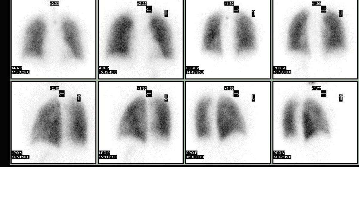 Matched V/Q Defects: Low Probability for PE
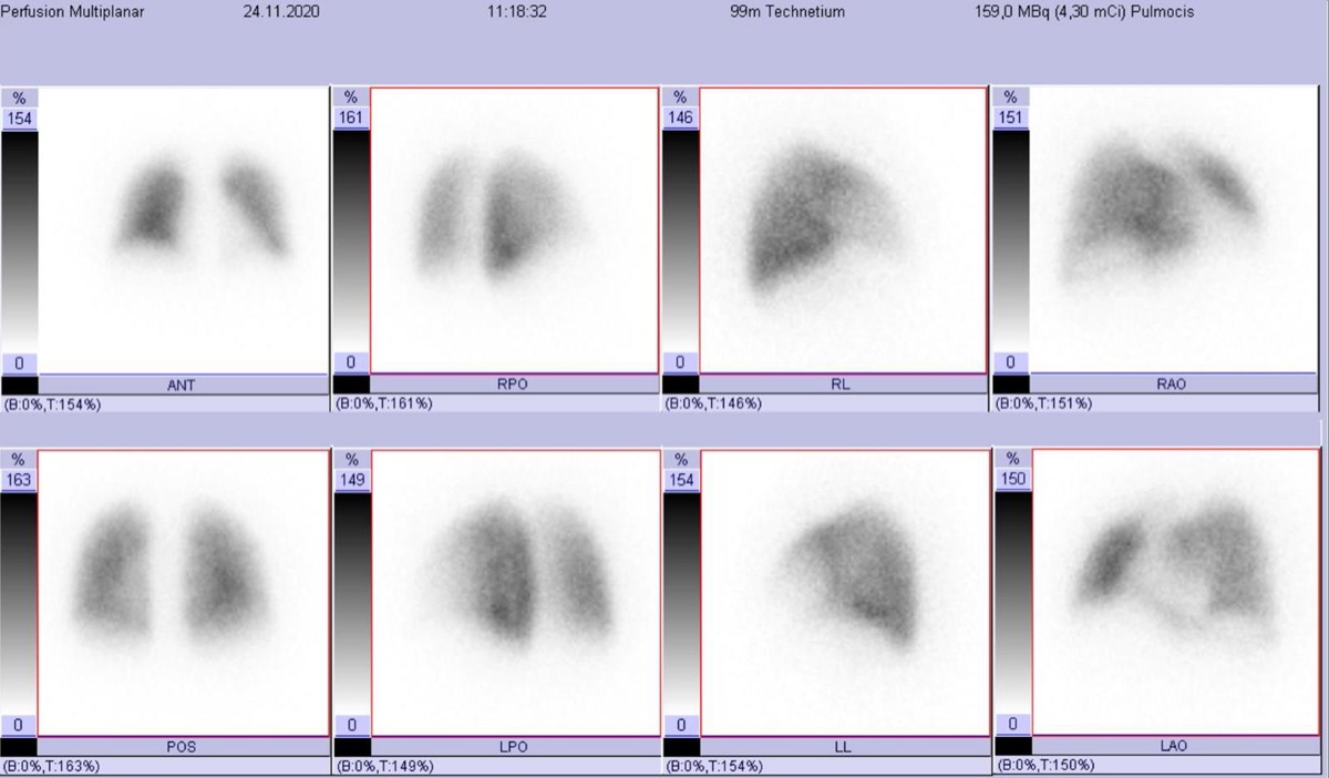 Normal Lung Perfusion Scan and Pulmonary Embolism
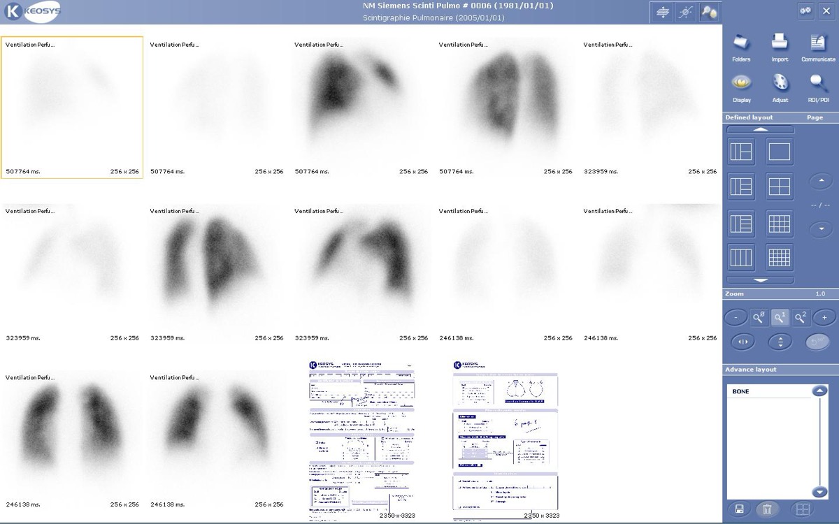 How Tc-99m MAA Lung Perfusion Imaging Works
Reducing MAA Particles in Pulmonary Hypertension
Stripe Sign on a Lung Perfusion Scan
V/Q Scan in COPD: Matched Defects
All 130 nuclear medicine images by category →
Practise image interpretation in the app
All 130 images as an interactive quiz, alongside 500 NMTCB and ARRT(N) questions with worked explanations, case studies and timed mock exams. 25 questions are free, no card and no account.