Matched V/Q Defects: Low Probability for PE
CC BY 3.0 · Myohan / Wikimedia Commons · source file
Question
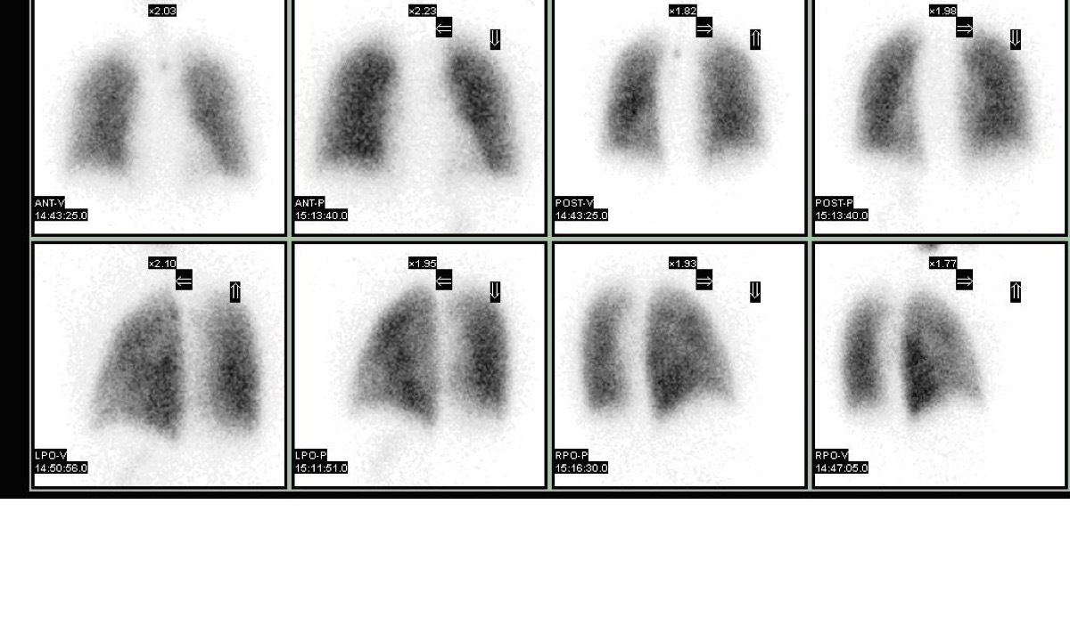
On a V/Q lung scan, both the ventilation and perfusion images show defects in the SAME region (a 'matched' defect). By PIOPED II criteria, a matched ventilation-perfusion defect in the setting of a normal or near-normal chest X-ray is most consistent with:
- AHigh probability for pulmonary embolism
- BLow probability for pulmonary embolism
- CCertain diagnosis of pulmonary embolism
- DNormal study
Show the answer and explanation
B. Low probability for pulmonary embolism
A matched V/Q defect — where both ventilation and perfusion are reduced in the same lung region — is characteristic of parenchymal lung disease (COPD, pneumonia, mucus plug), not PE. PE causes perfusion loss without ventilation loss (mismatch). According to PIOPED II criteria, matched defects with a normal or near-normal CXR are LOW probability for PE. Low probability does not mean zero risk — clinical probability must be integrated.
More lung v/q imaging images
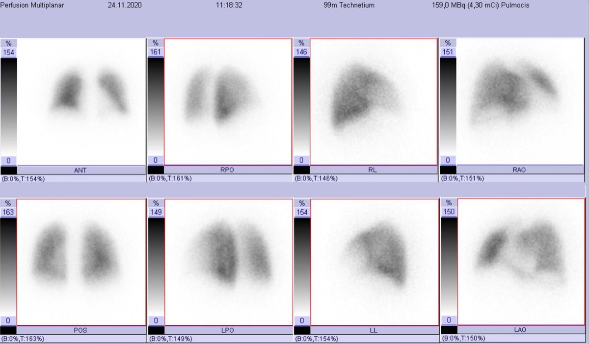 Normal Lung Perfusion Scan and Pulmonary Embolism
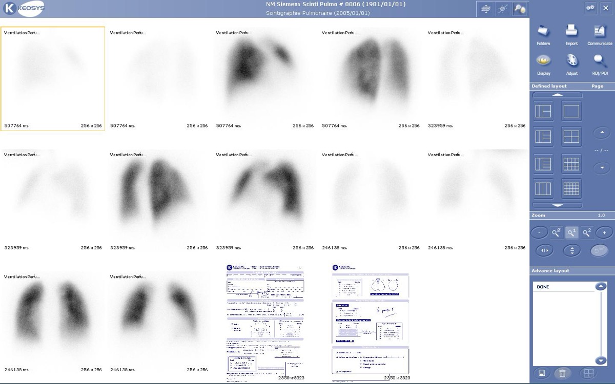 How Tc-99m MAA Lung Perfusion Imaging Works
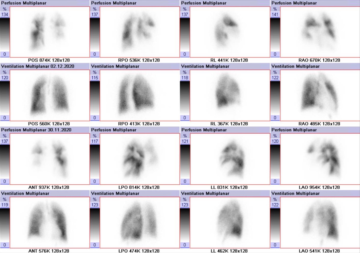 Reducing MAA Particles in Pulmonary Hypertension
Stripe Sign on a Lung Perfusion Scan
V/Q Scan in COPD: Matched Defects
V/Q Scan vs CTPA for Pulmonary Embolism in Pregnancy
All 130 nuclear medicine images by category →
Practise image interpretation in the app
All 130 images as an interactive quiz, alongside 500 NMTCB and ARRT(N) questions with worked explanations, case studies and timed mock exams. 25 questions are free, no card and no account.