Photopenic Bone Lesions: Multiple Myeloma
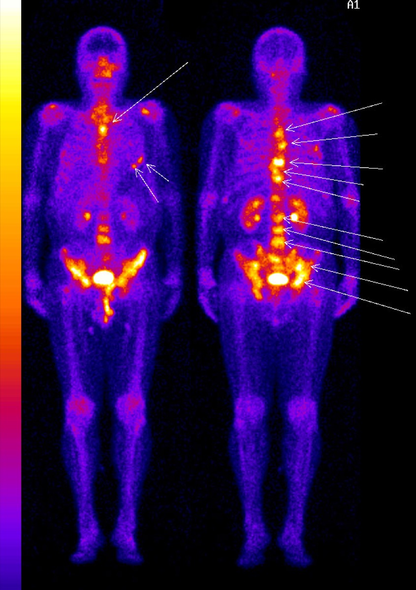
CC BY-SA 3.0 · Wikimedia Commons · source file
Question
A bone scan shows a focal area of DECREASED (photopenic or 'cold') uptake in the lumbar spine in a patient with known multiple myeloma. Why might myeloma lesions appear COLD on bone scan?
- AMultiple myeloma produces predominantly osteolytic lesions with little osteoblastic response
- BMultiple myeloma suppresses thyroid function, reducing MDP uptake
- CMyeloma lesions have increased blood flow that washes out the tracer
- DMultiple myeloma causes diffuse marrow infiltration that blocks tracer delivery
Show the answer and explanation
A. Multiple myeloma produces predominantly osteolytic lesions with little osteoblastic response
Multiple myeloma is a predominantly osteolytic malignancy — it destroys bone by stimulating osteoclasts without triggering a significant osteoblastic response. Since Tc-99m MDP localizes via osteoblastic activity (chemisorption onto hydroxyapatite during bone formation), purely lytic lesions with no osteoblastic response will show little or no tracer uptake — appearing cold (photopenic). This is why conventional bone scan has lower sensitivity for myeloma (~50–70%) compared to FDG PET/CT or whole-body MRI, which are preferred for myeloma staging.
More bone imaging images
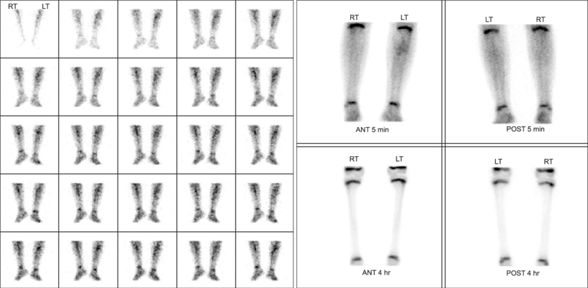 Cellulitis vs Osteomyelitis on Three-Phase Bone Scan
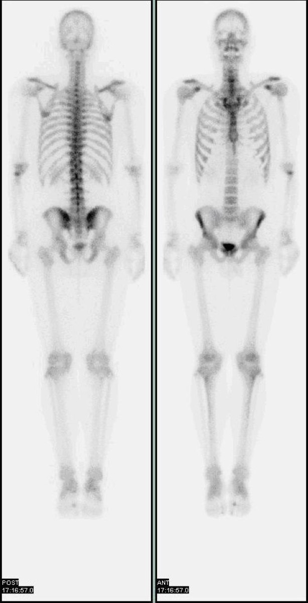 Normal Physiologic Uptake on a Bone Scan
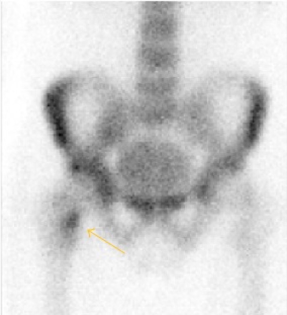 Shin Splints on Bone Scan: Medial Tibial Stress Syndrome
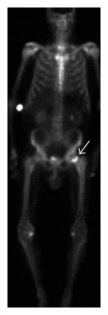 Avascular Necrosis of the Femoral Head on Bone Scan
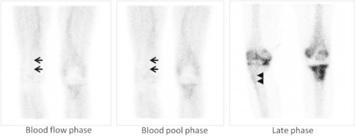 Flare Phenomenon on Bone Scan After Chemotherapy
 Bone Scan for Staging High-Risk Prostate Cancer
All 130 nuclear medicine images by category →
Practise image interpretation in the app
All 130 images as an interactive quiz, alongside 500 NMTCB and ARRT(N) questions with worked explanations, case studies and timed mock exams. 25 questions are free, no card and no account.