Paget’s Disease of Bone on Bone Scan

Public domain · Wikimedia Commons · source file
Question
A bone scan shows markedly intense, expanded-appearing uptake in the right hemipelvis and proximal femur, with bony enlargement visible on the delayed images. Alkaline phosphatase is markedly elevated. This pattern is most consistent with:
- AFibrous dysplasia
- BOsteoblastic metastases
- CAvascular necrosis
- DPaget's disease of bone
Show the answer and explanation
D. Paget's disease of bone
Paget's disease (osteitis deformans) produces dramatically intense radiotracer uptake in affected bones due to markedly increased bone turnover. The classic features on bone scan are: (1) expanded, enlarged bone contour; (2) intense homogeneous uptake (often described as 'hot as a light bulb'); (3) involvement extends to the articular surface; (4) markedly elevated alkaline phosphatase. The pelvis, skull, tibia, and vertebrae are the most common sites. Bone scan is more sensitive than radiographs for detecting Paget's disease and estimating extent of involvement.
More bone imaging images
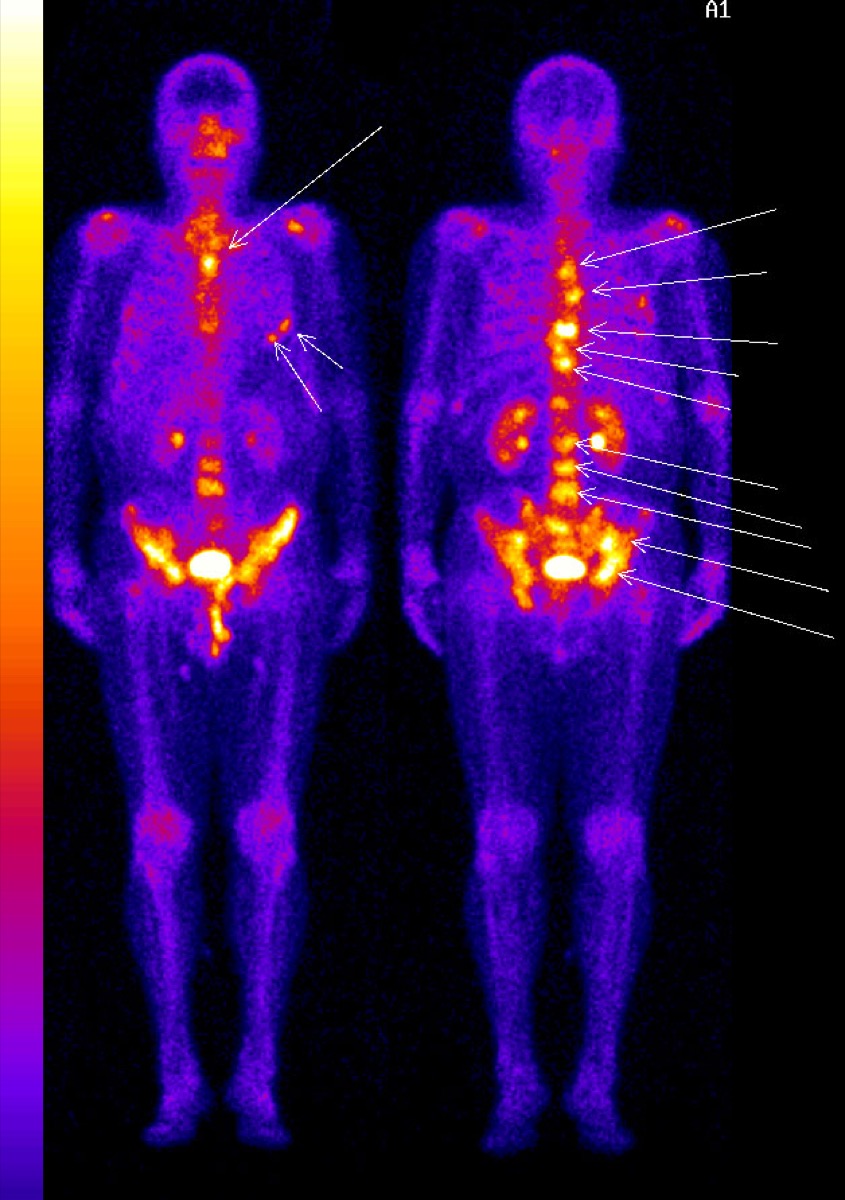 Photopenic Bone Lesions: Multiple Myeloma
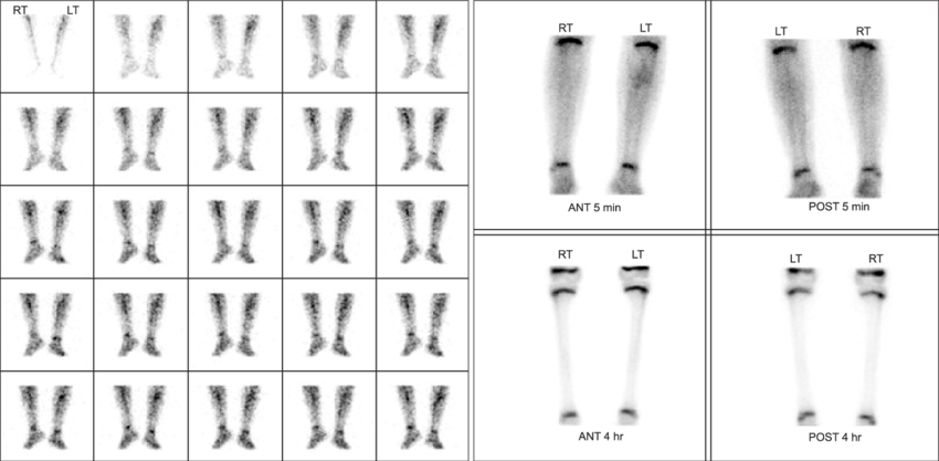 Cellulitis vs Osteomyelitis on Three-Phase Bone Scan
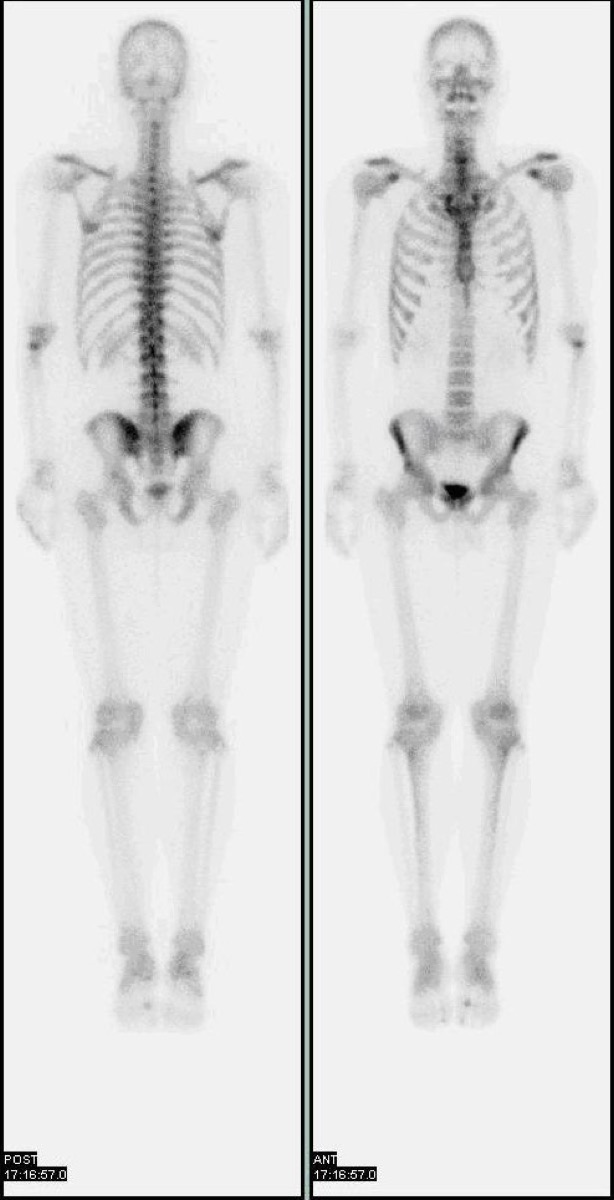 Normal Physiologic Uptake on a Bone Scan
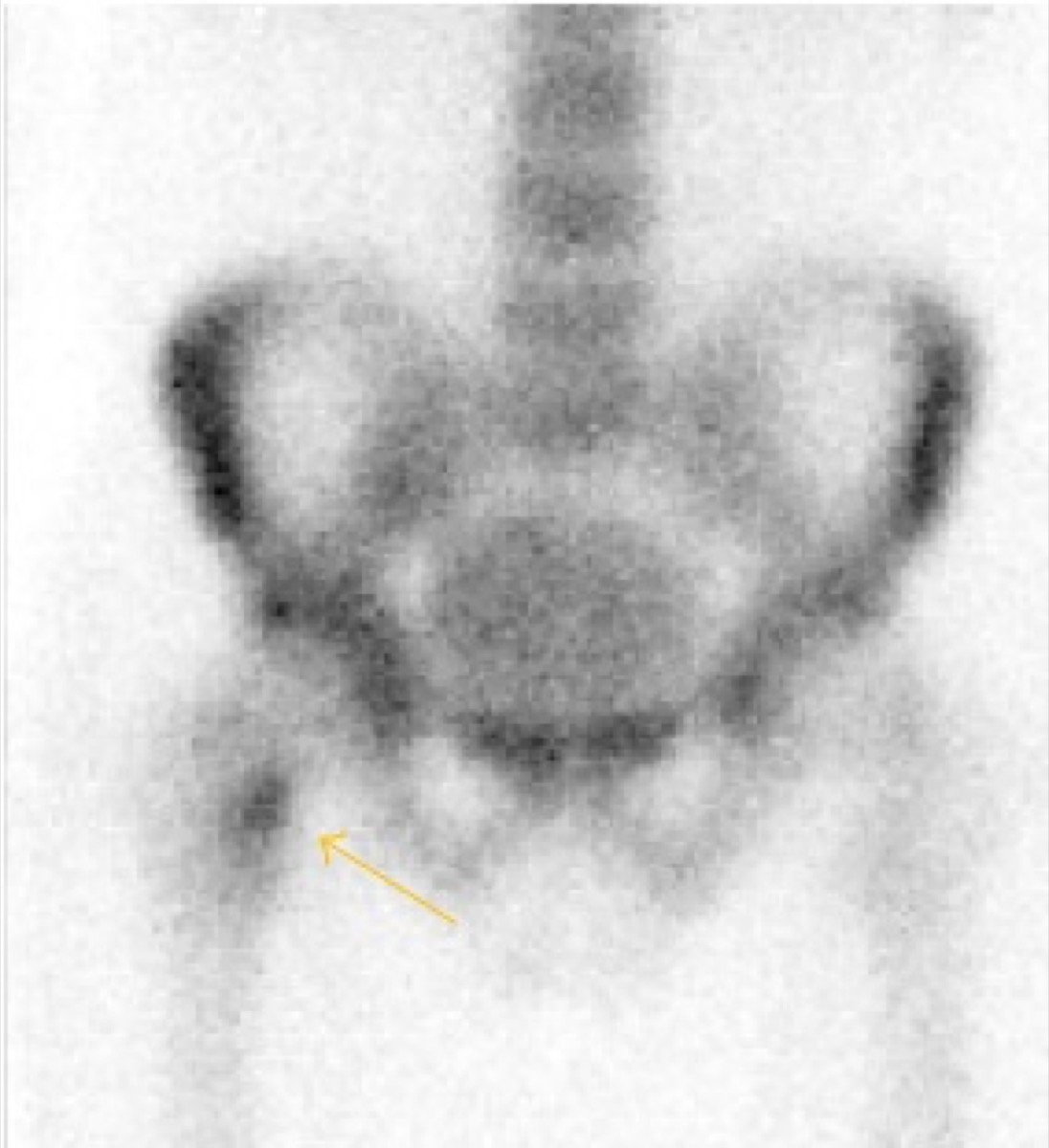 Shin Splints on Bone Scan: Medial Tibial Stress Syndrome
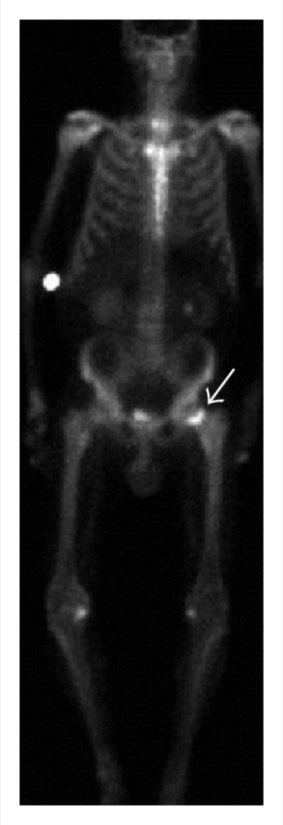 Avascular Necrosis of the Femoral Head on Bone Scan
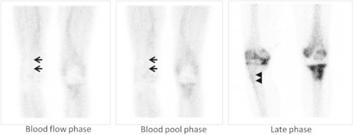 Flare Phenomenon on Bone Scan After Chemotherapy
All 130 nuclear medicine images by category →
Practise image interpretation in the app
All 130 images as an interactive quiz, alongside 500 NMTCB and ARRT(N) questions with worked explanations, case studies and timed mock exams. 25 questions are free, no card and no account.