Cellulitis vs Osteomyelitis on Three-Phase Bone Scan
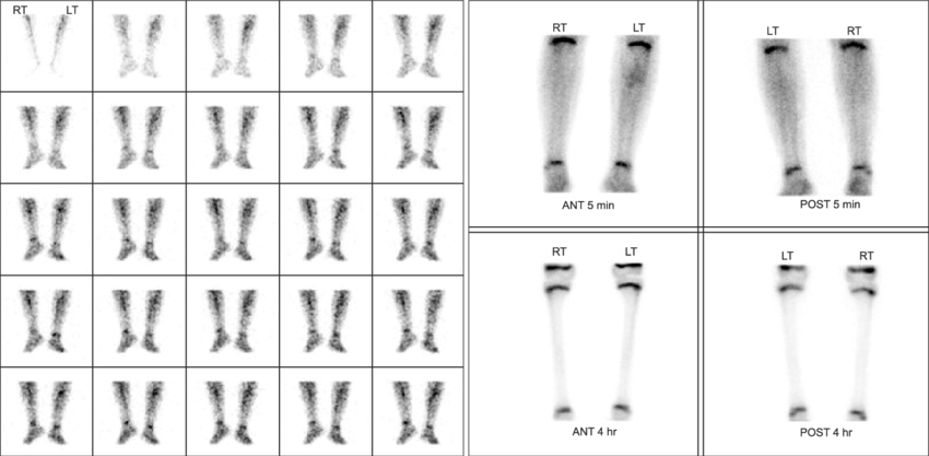
Provided clinical image
Question
A three-phase bone scan is performed in a diabetic patient with a red, swollen foot. The scan shows increased uptake in the FLOW and BLOOD POOL phases over the dorsal foot soft tissues, but the DELAYED phase shows normal bone uptake without focal abnormality. This pattern is most consistent with:
- AOsteomyelitis of the metatarsals
- BStress fracture of the fifth metatarsal
- CCRPS (Complex Regional Pain Syndrome)
- DCellulitis without underlying osteomyelitis
Show the answer and explanation
D. Cellulitis without underlying osteomyelitis
Cellulitis causes increased vascular activity (increased flow and blood pool phases) due to soft-tissue hyperemia, but WITHOUT increased delayed bone uptake, since the infection has not yet invaded bone. In contrast, OSTEOMYELITIS shows increased uptake in ALL THREE phases — flow, blood pool, AND delayed (bone) — because the infection directly involves the cortex and medullary bone. This distinction on three-phase bone scan is clinically critical in the diabetic foot, where distinguishing cellulitis from osteomyelitis guides antibiotic selection and surgical planning.
More bone imaging images
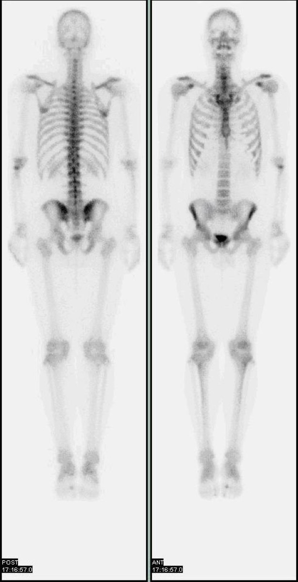 Normal Physiologic Uptake on a Bone Scan
 Shin Splints on Bone Scan: Medial Tibial Stress Syndrome
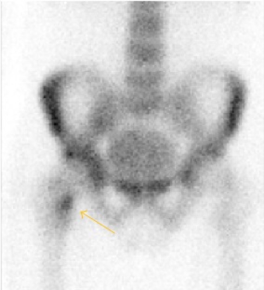
 Avascular Necrosis of the Femoral Head on Bone Scan
 Flare Phenomenon on Bone Scan After Chemotherapy
 Bone Scan for Staging High-Risk Prostate Cancer
 Fibrous Dysplasia on Bone Scan (Shepherd’s Crook)
All 130 nuclear medicine images by category →
Practise image interpretation in the app
All 130 images as an interactive quiz, alongside 500 NMTCB and ARRT(N) questions with worked explanations, case studies and timed mock exams. 25 questions are free, no card and no account.