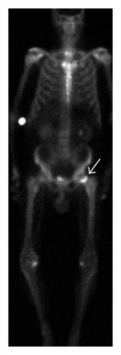
Superscan on Bone Scintigraphy
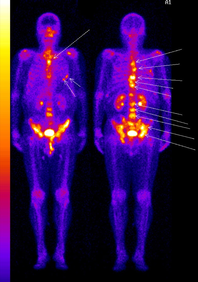
CC BY-SA 3.0 · Wikimedia Commons · source file
Question
A 'superscan' pattern on bone scintigraphy is characterized by diffusely increased skeletal uptake with absent or very faint kidney visualization. Which of the following is a common cause of a superscan?
- AWidespread skeletal metastases (especially prostate cancer) or metabolic bone disease
- BOsteoporosis
- CRenal failure with absent urine excretion
- DTechnically inadequate scan due to low administered activity
Show the answer and explanation
A. Widespread skeletal metastases (especially prostate cancer) or metabolic bone disease
A superscan occurs when there is such diffuse, intense skeletal uptake that virtually all radiotracer is extracted by bone, leaving little circulating tracer for renal excretion. The kidneys appear faint or absent, and the skeleton appears very bright. Common causes: (1) widespread osteoblastic skeletal metastases (especially prostate cancer); (2) metabolic bone disease (hyperparathyroidism, osteomalacia, renal osteodystrophy, Paget's disease). A superscan can be confused with a normal scan but is identified by the abnormally absent kidneys.
More bone imaging images
 Paget’s Disease of Bone on Bone Scan
Photopenic Bone Lesions: Multiple Myeloma
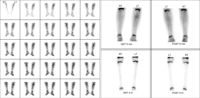 Cellulitis vs Osteomyelitis on Three-Phase Bone Scan
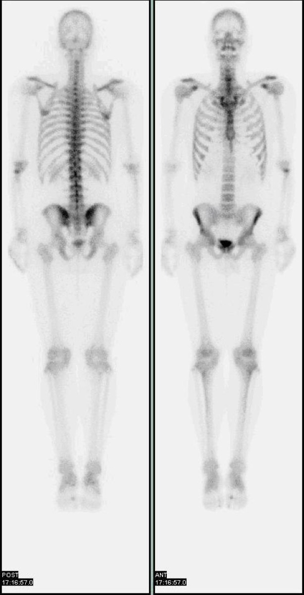 Normal Physiologic Uptake on a Bone Scan
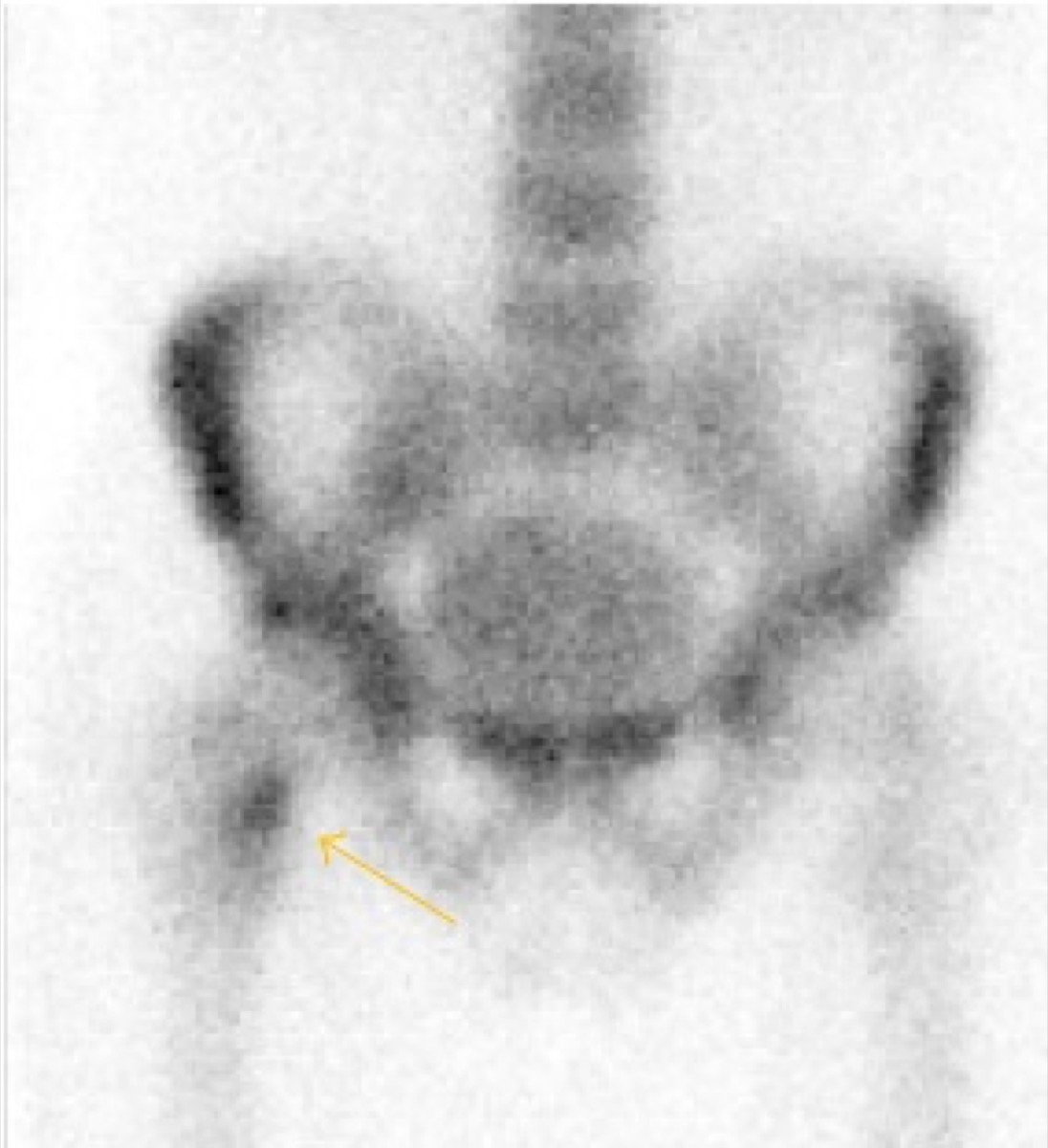 Shin Splints on Bone Scan: Medial Tibial Stress Syndrome
 Avascular Necrosis of the Femoral Head on Bone Scan
All 130 nuclear medicine images by category →
Practise image interpretation in the app
All 130 images as an interactive quiz, alongside 500 NMTCB and ARRT(N) questions with worked explanations, case studies and timed mock exams. 25 questions are free, no card and no account.