OctreoScan: Why Delayed 24-Hour Images
Provided clinical image
Question
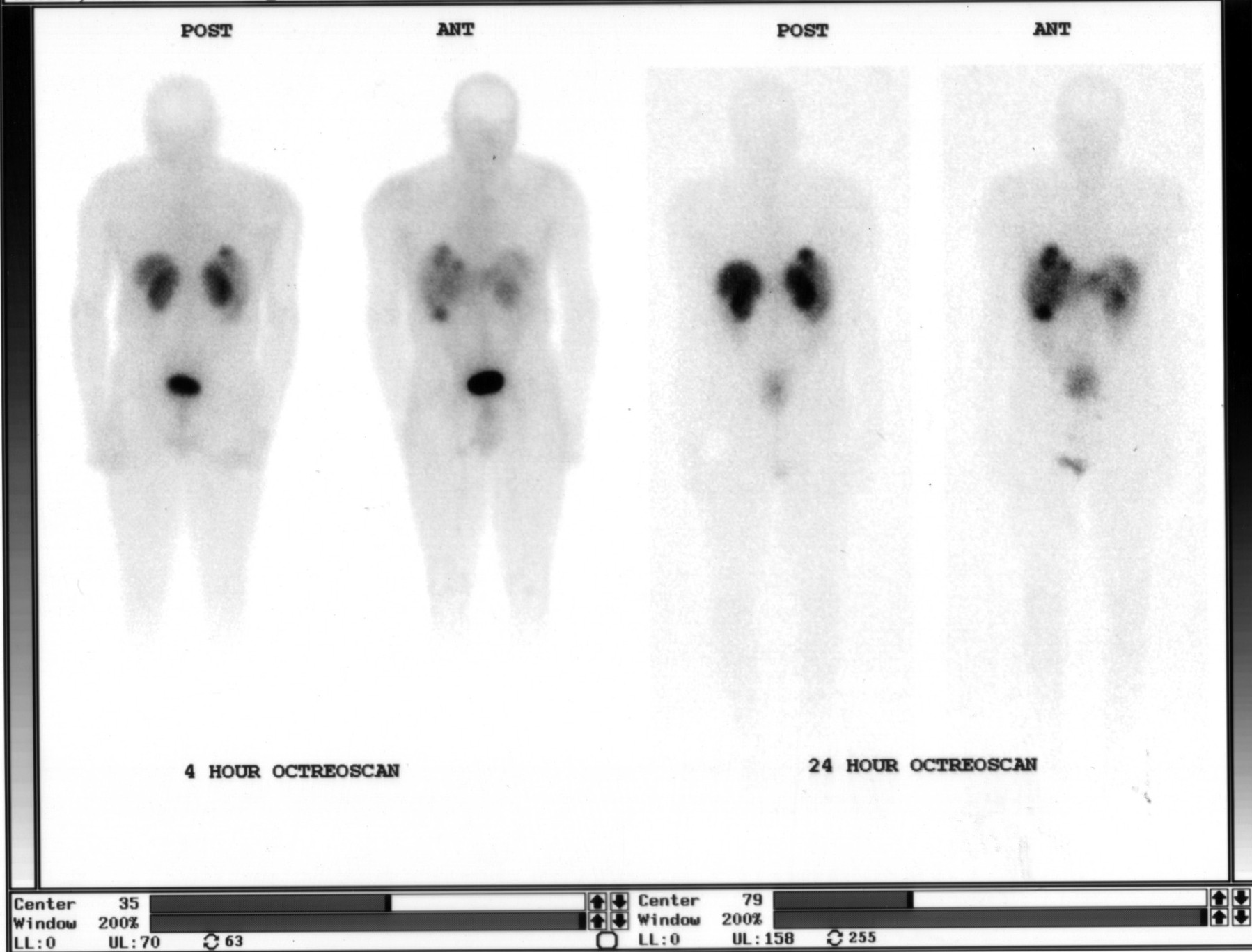
This Octreoscan was acquired at both 4 hours and 24 hours after injection. Why are delayed 24-hour images routinely obtained in addition to the 4-hour images?
- AAt 24 hours, the radiopharmaceutical reaches maximum uptake in neuroendocrine tumors, so only the delayed images are used for final interpretation
- BThe 4-hour images are used only for quality control to confirm proper injection technique and IV patency before proceeding to diagnostic imaging
- CBowel activity from hepatobiliary excretion can obscure abdominal lesions on early images, and the 24-hour delay allows this activity to clear for better lesion visibility
- DRegulatory guidelines require dual-timepoint imaging for all In-111 labeled agents to document biodistribution at two distinct phases
Show the answer and explanation
C. Bowel activity from hepatobiliary excretion can obscure abdominal lesions on early images, and the 24-hour delay allows this activity to clear for better lesion visibility
In-111 pentetreotide is excreted via the hepatobiliary system into the bowel, which can appear as focal abdominal activity on early (4-hour) images and potentially obscure true lesions. Acquiring 24-hour delayed images allows this bowel activity to clear, significantly improving the ability to identify and confirm abdominal and pelvic lesions.
More pet and oncology images
OctreoScan Liver Uptake: Somatostatin Receptors
 Normal FDG PET Biodistribution
 OctreoScan (In-111 Octreotide): How It Localizes
 I-123 MIBG Uptake Mechanism in Pheochromocytoma
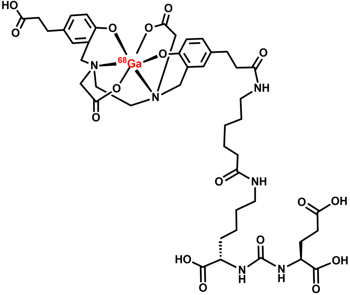 Ga-68 PSMA PET/CT in Prostate Cancer
Ga-68 DOTATATE PET vs OctreoScan for Neuroendocrine Tumors
All 130 nuclear medicine images by category →
Practise image interpretation in the app
All 130 images as an interactive quiz, alongside 500 NMTCB and ARRT(N) questions with worked explanations, case studies and timed mock exams. 25 questions are free, no card and no account.