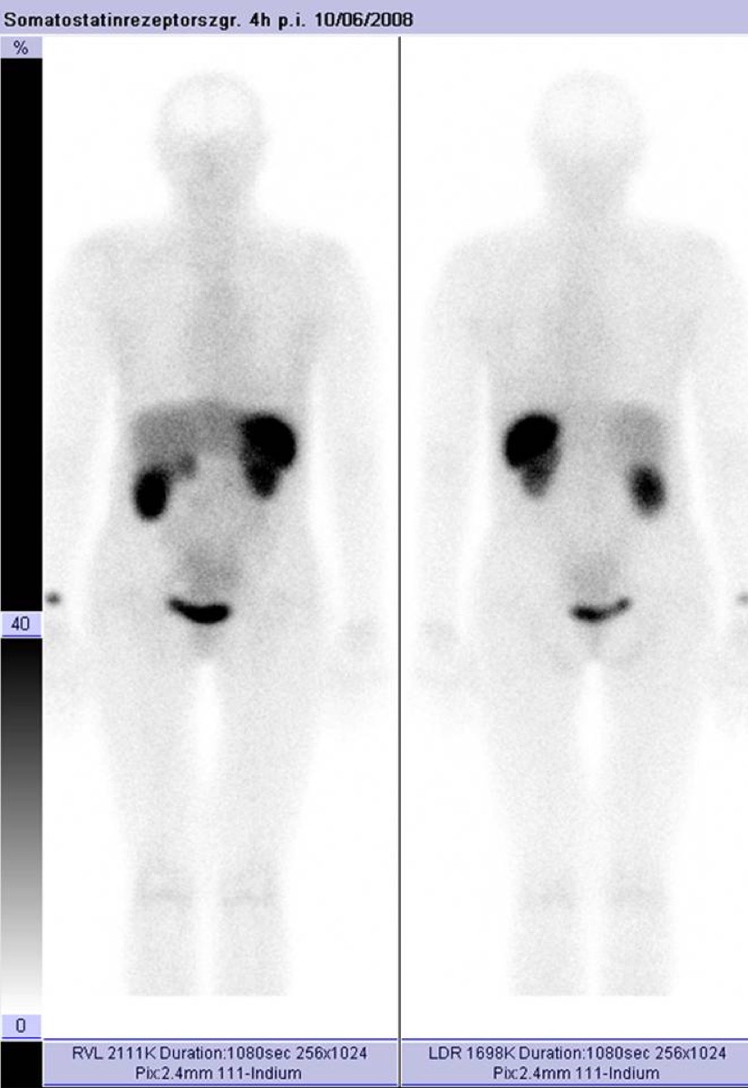
Carcinoid Tumor on Somatostatin Receptor Imaging
CC BY-SA 3.0 · Wikimedia Commons · source file
Question
A patient with diarrhea, flushing, and an elevated 24-hour urine 5-HIAA is found to have a small bowel mass on CT. Ga-68 DOTATATE PET/CT shows intense SSTR-avid uptake in the primary tumor and liver metastases. What is the MOST likely diagnosis?
- AInsulinoma — always somatostatin receptor negative
- BGastrointestinal adenocarcinoma
- CLymphoma of the small bowel
- DCarcinoid tumor (well-differentiated serotonin-secreting NET) — the classic presentation of carcinoid syndrome with positive somatostatin receptor imaging
Show the answer and explanation
D. Carcinoid tumor (well-differentiated serotonin-secreting NET) — the classic presentation of carcinoid syndrome with positive somatostatin receptor imaging
The clinical triad of diarrhea, flushing, and elevated 5-HIAA (5-hydroxyindoleacetic acid, a serotonin metabolite) in combination with a small bowel mass and liver metastases is CLASSIC for carcinoid syndrome caused by a midgut carcinoid tumor (well-differentiated serotonin-secreting NET). Carcinoid syndrome only occurs when metastases reach the liver (bypassing first-pass serotonin metabolism by the liver). Well-differentiated NETs (carcinoids) overexpress SSTR2 and are strongly positive on Ga-68 DOTATATE PET/CT. SSTR imaging is used for staging, treatment selection (PRRT eligibility), and monitoring. 5-HIAA is the classic biochemical marker.
More pet and oncology images
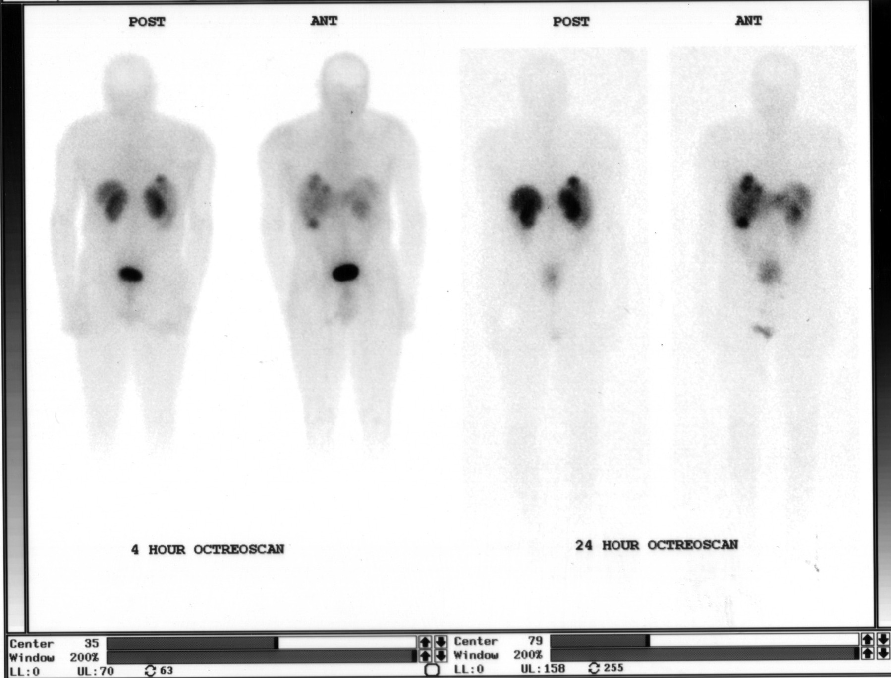 OctreoScan: Why Delayed 24-Hour Images
OctreoScan Liver Uptake: Somatostatin Receptors
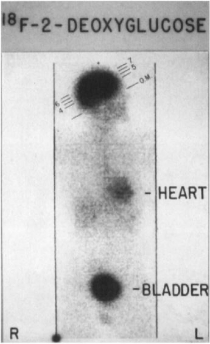 Normal FDG PET Biodistribution
OctreoScan (In-111 Octreotide): How It Localizes
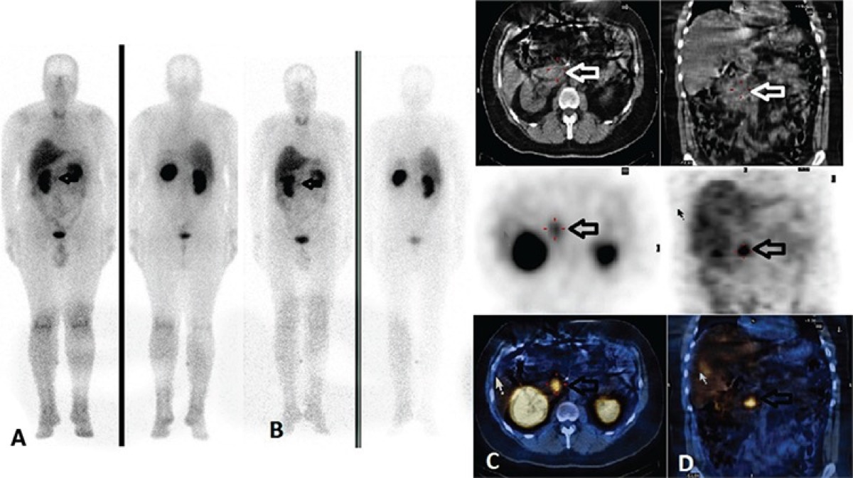 I-123 MIBG Uptake Mechanism in Pheochromocytoma
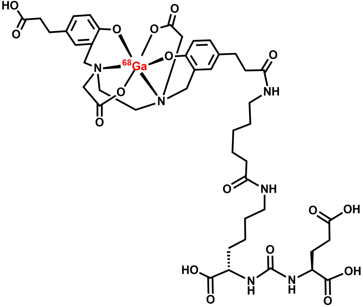 Ga-68 PSMA PET/CT in Prostate Cancer
All 130 nuclear medicine images by category →
Practise image interpretation in the app
All 130 images as an interactive quiz, alongside 500 NMTCB and ARRT(N) questions with worked explanations, case studies and timed mock exams. 25 questions are free, no card and no account.