I-123 MIBG Uptake Mechanism in Pheochromocytoma
CC BY 4.0 · Wikimedia Commons · source file
Question
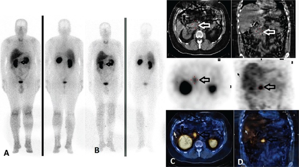
This MIBG scan shows intense focal uptake in the right adrenal region in a patient with hypertensive crises. MIBG localizes to chromaffin tissue by what mechanism, making it useful for detecting pheochromocytoma?
- APassive diffusion across the adrenal cell membrane proportional to regional blood flow, with intracellular trapping by mitochondrial binding proteins in steroidogenic cells
- BReceptor-mediated endocytosis via glucocorticoid receptors on the adrenal cortex, with retrograde axonal transport to the sympathetic ganglia
- CActive uptake via the norepinephrine transporter (NET) into chromaffin cells, the same pathway used by catecholamines
- DFDG-like accumulation driven by upregulated hexokinase activity and GLUT overexpression in hyperfunctioning adrenal medullary tissue
Show the answer and explanation
C. Active uptake via the norepinephrine transporter (NET) into chromaffin cells, the same pathway used by catecholamines
MIBG (metaiodobenzylguanidine) is a structural analogue of norepinephrine. It is taken up by chromaffin cells (adrenal medulla, sympathetic nerve terminals) via the norepinephrine transporter (NET) and stored in secretory granules. Pheochromocytomas and paragangliomas (catecholamine-secreting tumors of chromaffin tissue) overexpress NET and accumulate MIBG intensely. MIBG is also used for neuroblastoma imaging and therapy (I-131 MIBG). Certain medications that block NET (tricyclic antidepressants, labetalol, calcium channel blockers) must be stopped before MIBG scanning.
More pet and oncology images
 Ga-68 PSMA PET/CT in Prostate Cancer
 Ga-68 DOTATATE PET vs OctreoScan for Neuroendocrine Tumors
 Deauville Score 1–2: Complete Metabolic Response
FDG-Avid Benign Disease: Infection and Inflammation Mimics
How SUV Is Calculated on FDG PET
FDG PET/CT Staging of Non-Small Cell Lung Cancer
All 130 nuclear medicine images by category →
Practise image interpretation in the app
All 130 images as an interactive quiz, alongside 500 NMTCB and ARRT(N) questions with worked explanations, case studies and timed mock exams. 25 questions are free, no card and no account.