Normal FDG PET Biodistribution
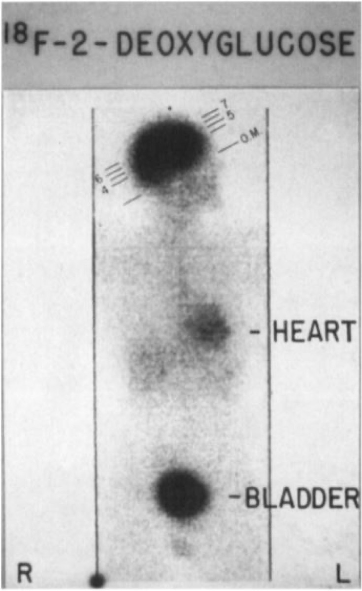
CC BY-SA 4.0 · Pennradiology / Wikimedia Commons · source file
Question
This FDG PET scan shows the normal distribution of F-18 fluorodeoxyglucose. Which of the following structures has HIGH FDG uptake in a normal study?
- ALiver only
- BBrain, myocardium, liver, and urinary tract
- CBone marrow only
- DLung and muscle exclusively
Show the answer and explanation
B. Brain, myocardium, liver, and urinary tract
Normal FDG distribution includes: (1) BRAIN — highest uptake due to obligatory glucose dependence; (2) MYOCARDIUM — variable uptake depending on fasting state (high in fed state, suppressed in fasted); (3) LIVER — moderate uniform uptake; (4) URINARY TRACT (kidneys, ureters, bladder) — FDG is excreted unchanged in urine; (5) SPLEEN — mild uptake. Skeletal muscle uptake is low at rest but increases significantly with exercise. Recognizing normal FDG distribution is critical to avoid misinterpreting physiologic uptake as pathology.
More pet and oncology images
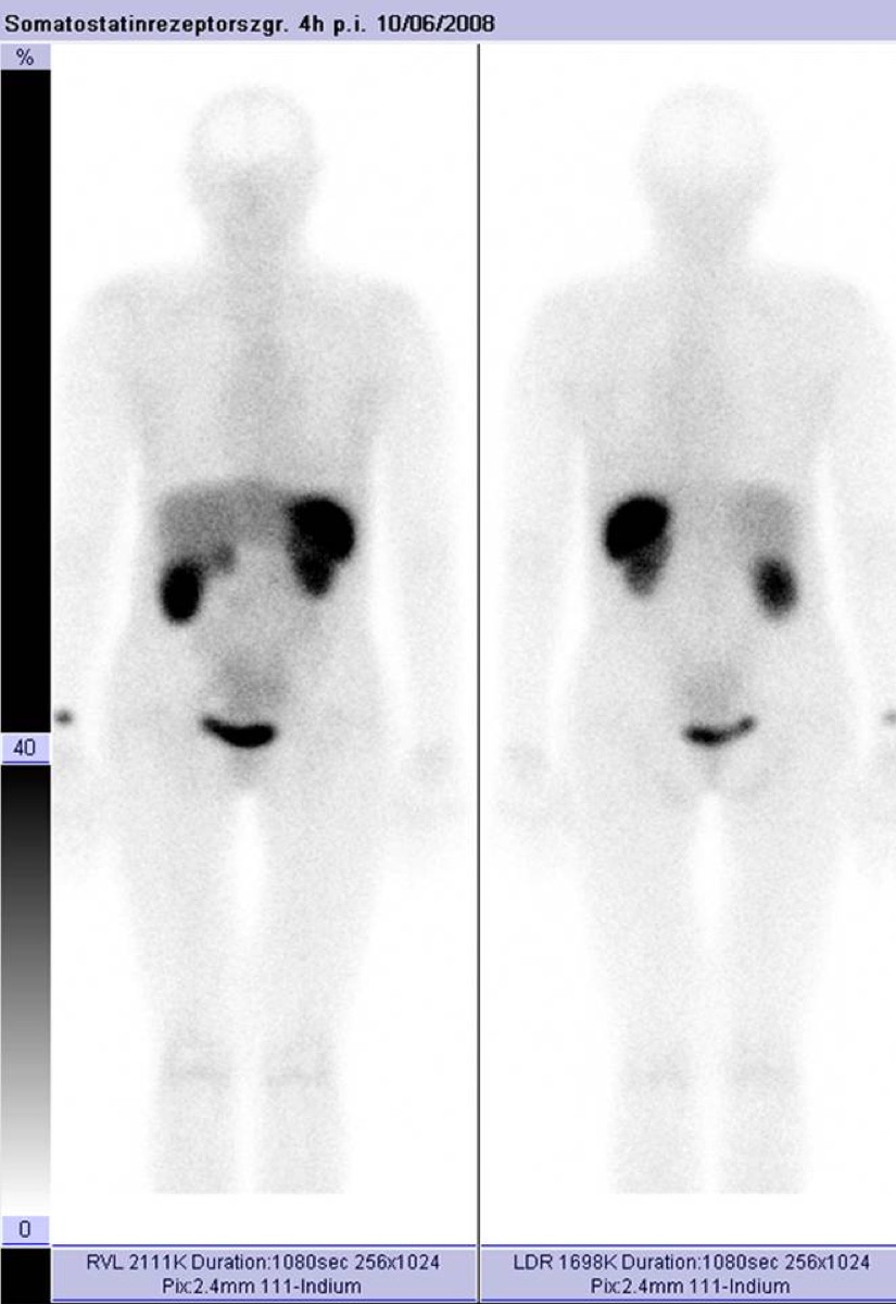 OctreoScan (In-111 Octreotide): How It Localizes
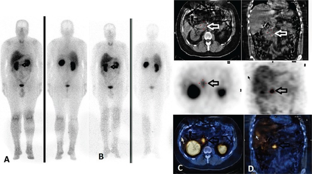 I-123 MIBG Uptake Mechanism in Pheochromocytoma
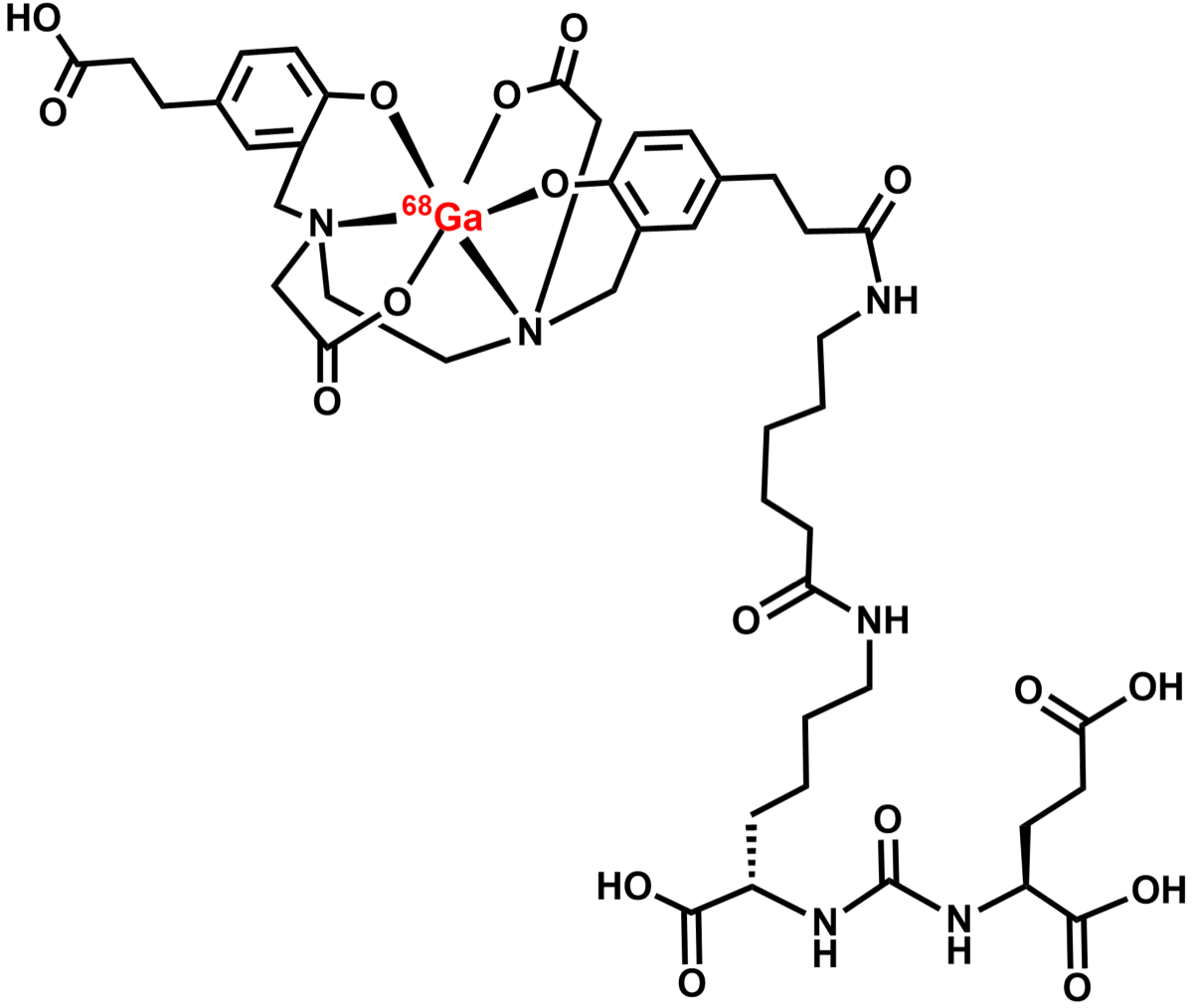 Ga-68 PSMA PET/CT in Prostate Cancer
Ga-68 DOTATATE PET vs OctreoScan for Neuroendocrine Tumors
Deauville Score 1–2: Complete Metabolic Response
FDG-Avid Benign Disease: Infection and Inflammation Mimics
All 130 nuclear medicine images by category →
Practise image interpretation in the app
All 130 images as an interactive quiz, alongside 500 NMTCB and ARRT(N) questions with worked explanations, case studies and timed mock exams. 25 questions are free, no card and no account.