Ga-68 PSMA PET/CT in Prostate Cancer
CC BY-SA 4.0 · Wikimedia Commons · source file
Question
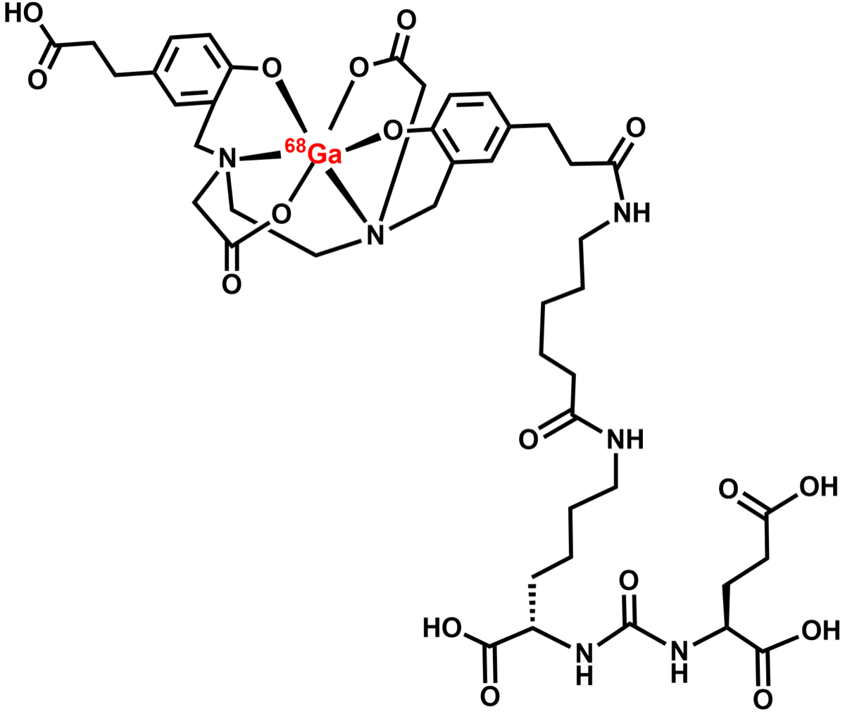
This Ga-68 PSMA PET/CT shows abnormal foci of PSMA-avid uptake. PSMA (prostate-specific membrane antigen) PET is the preferred imaging modality for which of the following clinical scenarios?
- AInitial staging of breast cancer, particularly in patients with HER2-positive disease where standard CT underestimates axillary nodal involvement and distant metastatic burden
- BAssessment of cardiac viability in ischemic cardiomyopathy, where PSMA expression on cardiac fibroblasts correlates with myocardial fibrosis and remodeling after infarction
- CBiochemical recurrence and initial staging of prostate cancer
- DDetection of pulmonary nodules in high-risk lung cancer screening populations, where PSMA expression on tumor neovasculature provides superior sensitivity to low-dose CT
Show the answer and explanation
C. Biochemical recurrence and initial staging of prostate cancer
Ga-68 PSMA-11 PET/CT targets prostate-specific membrane antigen, a glycoprotein overexpressed on prostate cancer cells. It is FDA-approved for initial staging of high-risk prostate cancer and for evaluation of biochemical recurrence (rising PSA after treatment). PSMA PET detects recurrent/metastatic prostate cancer at much lower PSA levels than conventional imaging (CT/bone scan). Normal PSMA uptake is seen in kidneys, liver, salivary glands, spleen, and small bowel.
More pet and oncology images
 Ga-68 DOTATATE PET vs OctreoScan for Neuroendocrine Tumors
 Deauville Score 1–2: Complete Metabolic Response
FDG-Avid Benign Disease: Infection and Inflammation Mimics
How SUV Is Calculated on FDG PET
FDG PET/CT Staging of Non-Small Cell Lung Cancer
Brown Fat Uptake on FDG PET
All 130 nuclear medicine images by category →
Practise image interpretation in the app
All 130 images as an interactive quiz, alongside 500 NMTCB and ARRT(N) questions with worked explanations, case studies and timed mock exams. 25 questions are free, no card and no account.