Furosemide Dose for Diuretic Renography
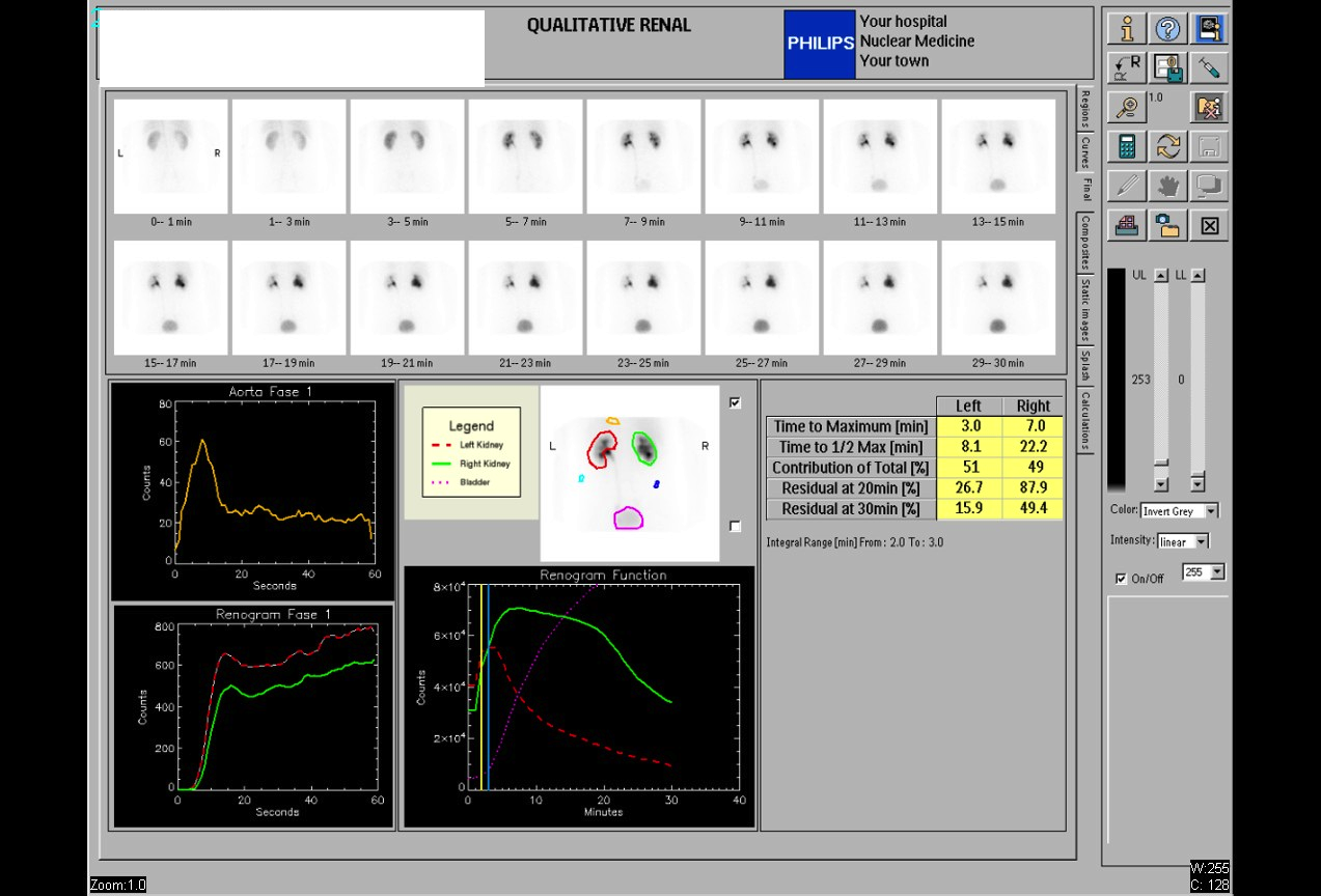
Provided clinical image
Question
During a diuretic renal scan (MAG3 with Lasix), furosemide is injected to help determine whether retained tracer in the collecting system is due to true obstruction or just a dilated but non-obstructed system. As the nuclear medicine technologist performing this study, what is the STANDARD adult dose and route of administration for furosemide?
- A20 mg administered orally 1 hour before the scan begins, to ensure adequate diuresis during the imaging period
- B40 mg (0.5 mg/kg) administered intravenously during the scan, typically at 20 minutes post-injection or when the collecting system is well-visualized
- C100 mg administered intramuscularly at the start of the scan, allowing gradual absorption to produce sustained diuresis throughout imaging
- D10 mg administered as a subcutaneous injection 30 minutes before the radiopharmaceutical, to pre-hydrate the collecting system prior to tracer arrival
Show the answer and explanation
B. 40 mg (0.5 mg/kg) administered intravenously during the scan, typically at 20 minutes post-injection or when the collecting system is well-visualized
The standard furosemide (Lasix) dose for a diuretic renogram is 0.5 mg/kg IV (maximum 40 mg for adults). It is administered intravenously — either at the start of the study (F+0 protocol), 15–20 minutes into the scan (F+20 protocol, most common), or 15 minutes before the radiopharmaceutical (F-15 protocol). The IV route ensures rapid onset of diuresis. The resulting increased urine flow will wash out tracer from a non-obstructed dilated system (T½ <10 min post-Lasix) but will fail to clear a truly obstructed system (T½ >20 min post-Lasix).
More renal and genitourinary images
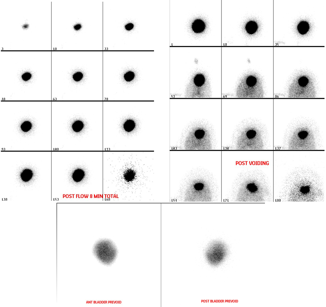 Direct Radionuclide Cystogram Technique
Vesicoureteral Reflux on Radionuclide Cystogram
Why Post-Void Images on a Radionuclide Cystogram
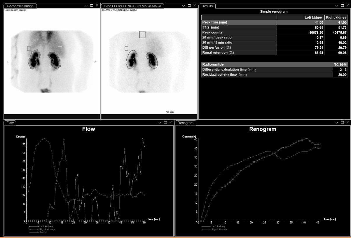 Differential Renal Function on MAG3
Bilateral Impaired Drainage on MAG3 Renogram
Motion Correction in MAG3 Renal Scans
All 130 nuclear medicine images by category →
Practise image interpretation in the app
All 130 images as an interactive quiz, alongside 500 NMTCB and ARRT(N) questions with worked explanations, case studies and timed mock exams. 25 questions are free, no card and no account.