Why MAG3 for Renal Imaging: Tubular Secretion
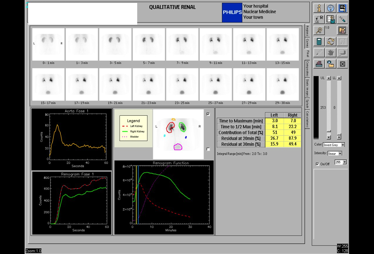
Provided clinical image
Question
The sequential images shown are from a qualitative renal scan used to evaluate kidney drainage. Looking at the renogram curves, the left kidney (red dashed line) peaks early and clears normally, while the right kidney (green line) peaks later and retains tracer through 30 minutes. Which radiopharmaceutical is MOST COMMONLY used for this type of renal drainage study?
- ATc-99m DMSA (dimercaptosuccinic acid) — binds to renal cortical tubular cells and provides static images of cortical function
- BTc-99m MDP (methylene diphosphonate) — taken up by osteoblasts and used primarily for renal cortical scanning due to urinary excretion
- CTc-99m MAG3 (mercaptoacetyltriglycine) — excreted by tubular secretion, providing dynamic images ideal for evaluating renal blood flow and drainage
- DTc-99m sulfur colloid — taken up by the reticuloendothelial system and used for renal drainage studies when MAG3 is unavailable
Show the answer and explanation
C. Tc-99m MAG3 (mercaptoacetyltriglycine) — excreted by tubular secretion, providing dynamic images ideal for evaluating renal blood flow and drainage
Tc-99m MAG3 (mercaptoacetyltriglycine) is the most commonly used radiopharmaceutical for dynamic renal studies (renography). It is excreted primarily by tubular secretion (not glomerular filtration), giving it a high extraction efficiency — especially useful in patients with reduced renal function. MAG3 produces high-quality renogram curves that allow evaluation of renal perfusion, cortical transit, and collecting system drainage. Tc-99m DMSA is used for static cortical imaging (scarring, pyelonephritis) and does not evaluate drainage.
More renal and genitourinary images
Furosemide Dose for Diuretic Renography
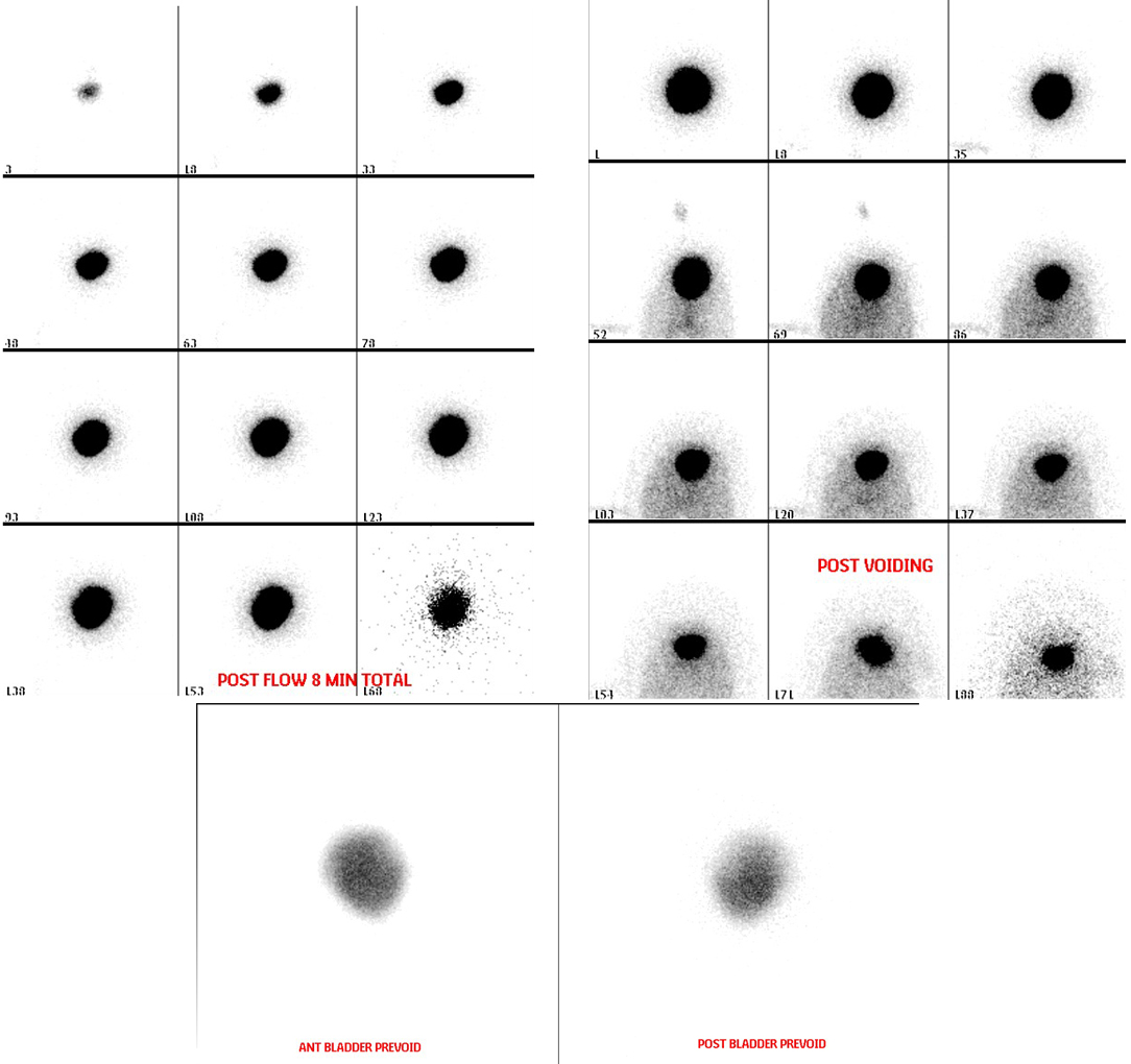 Direct Radionuclide Cystogram Technique
Vesicoureteral Reflux on Radionuclide Cystogram
Why Post-Void Images on a Radionuclide Cystogram
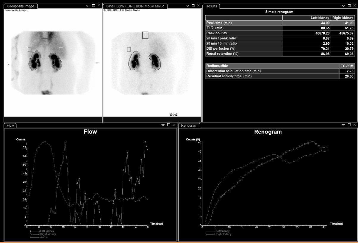 Differential Renal Function on MAG3
Bilateral Impaired Drainage on MAG3 Renogram
All 130 nuclear medicine images by category →
Practise image interpretation in the app
All 130 images as an interactive quiz, alongside 500 NMTCB and ARRT(N) questions with worked explanations, case studies and timed mock exams. 25 questions are free, no card and no account.