Prone Imaging in Cardiac SPECT
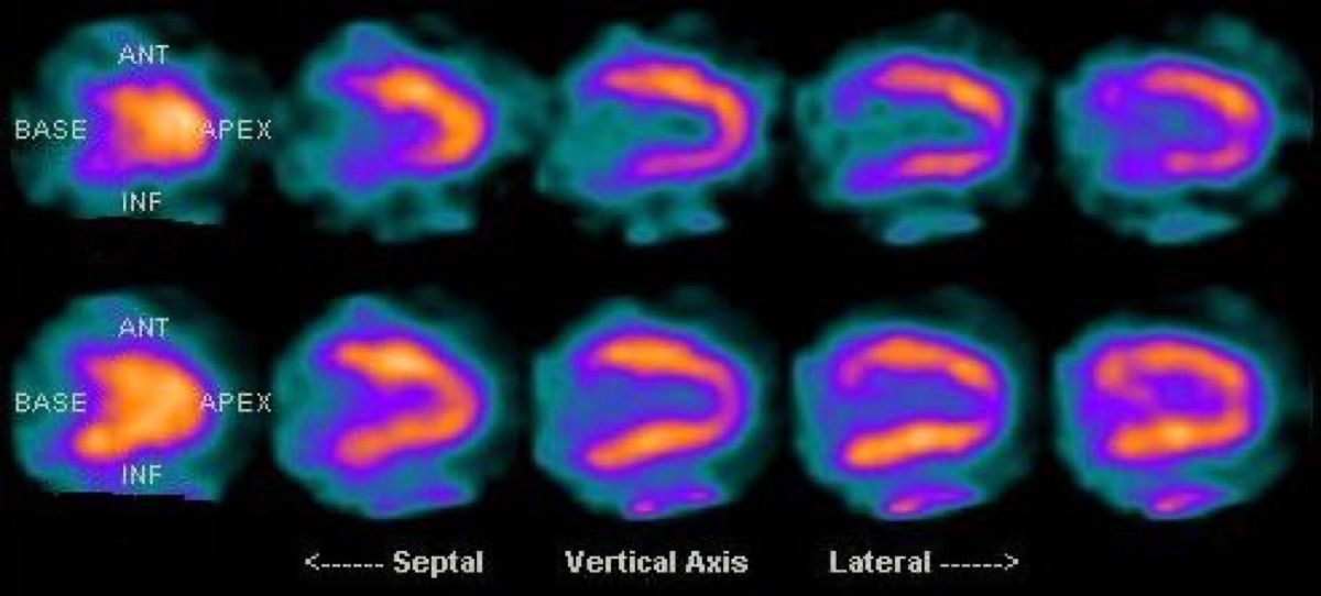
Public domain · KieranMaher / Wikimedia Commons · source file
Question
In addition to CT attenuation correction, prone (face-down) imaging is sometimes added to supine cardiac SPECT. The PRIMARY purpose of prone imaging in cardiac SPECT is:
- ATo reduce inferior and inferolateral wall attenuation artifacts caused by the diaphragm
- BTo improve detection of right ventricular perfusion defects
- CTo reduce patient motion during acquisition
- DTo increase administered activity to the heart
Show the answer and explanation
A. To reduce inferior and inferolateral wall attenuation artifacts caused by the diaphragm
Prone imaging is performed by placing the patient face-down, which causes the diaphragm and abdominal contents to fall away from the inferior wall of the heart due to gravity. This reduces the diaphragmatic attenuation that commonly creates artifactual inferior wall perfusion defects on supine imaging. An inferior wall defect that DISAPPEARS on prone imaging is almost certainly a diaphragmatic attenuation artifact. An inferior wall defect that PERSISTS on both supine and prone images is more likely to represent true RCA territory ischemia or infarction. Prone imaging is particularly useful when CT attenuation correction is not available.
More cardiac imaging images
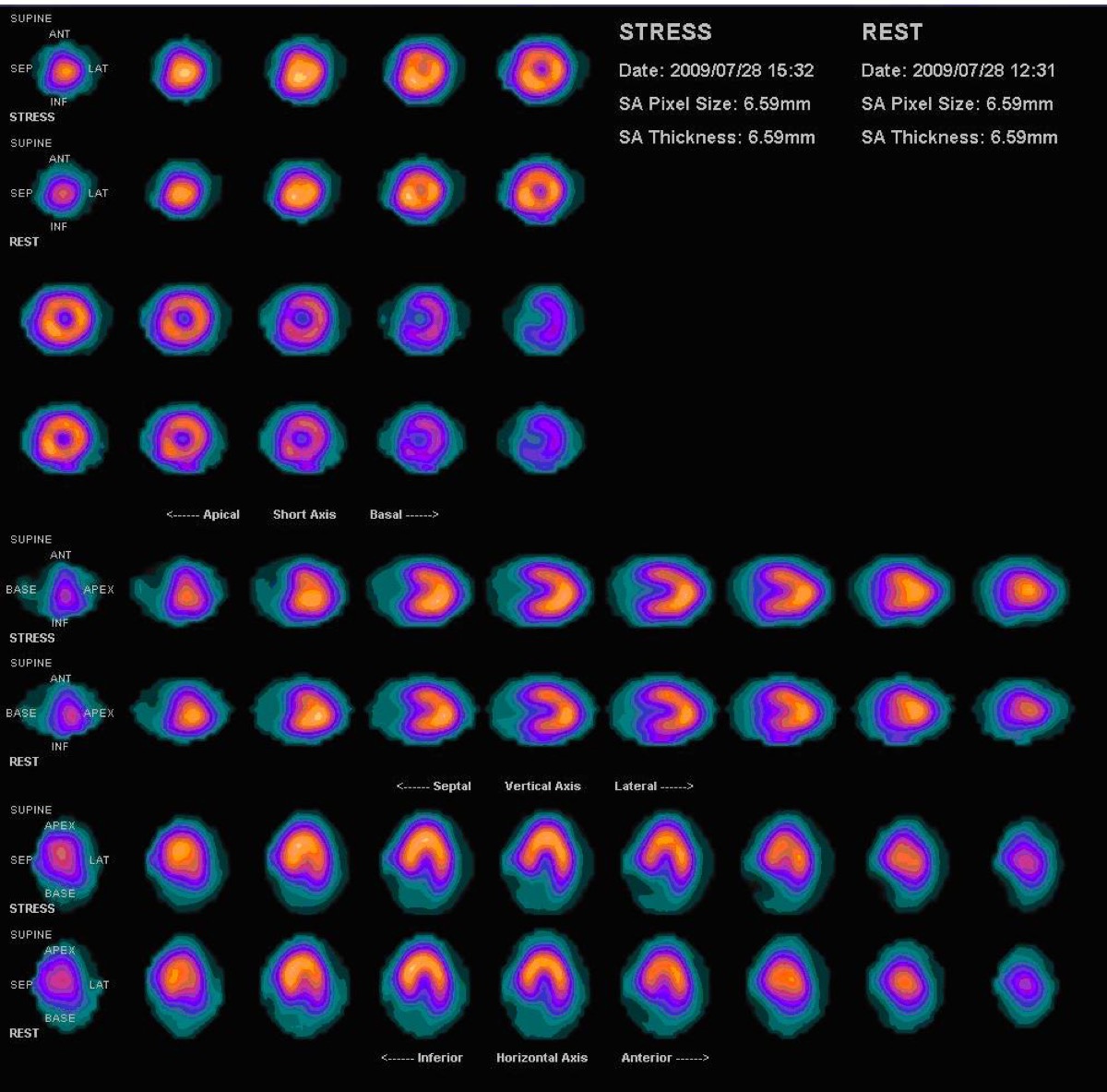 Prominent Right Ventricle on MPI: RV Hypertrophy
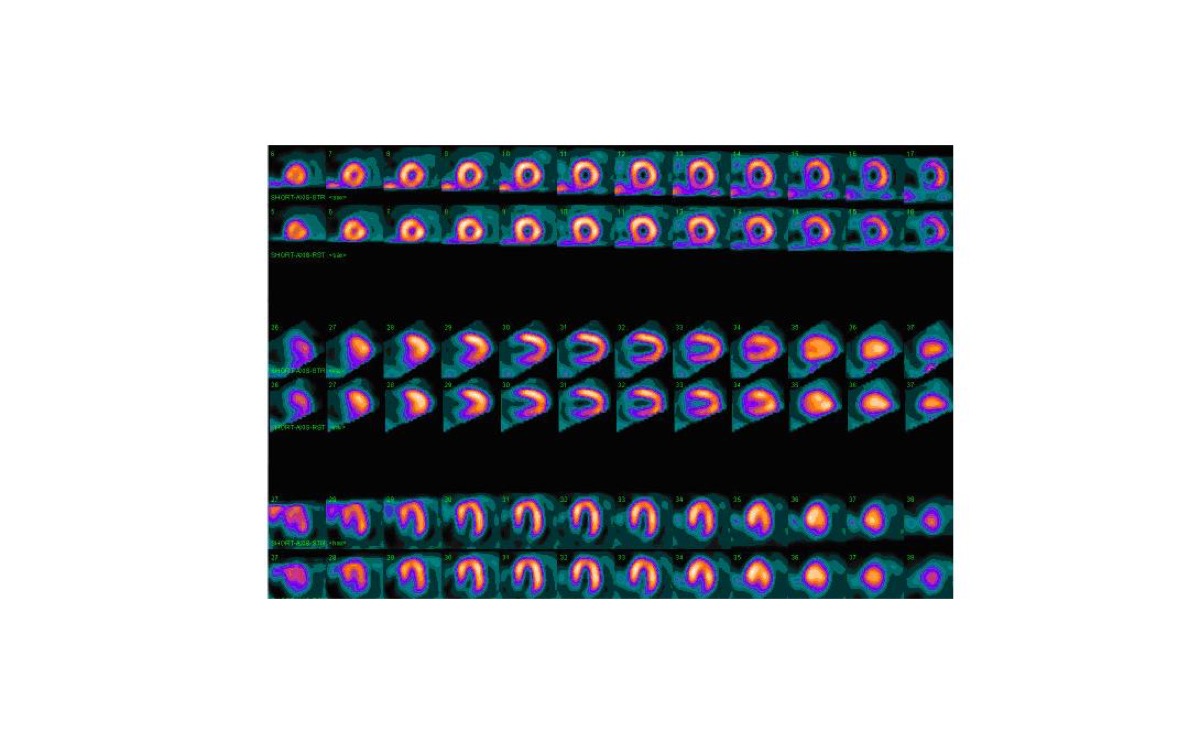 Dobutamine Stress for Myocardial Perfusion Imaging
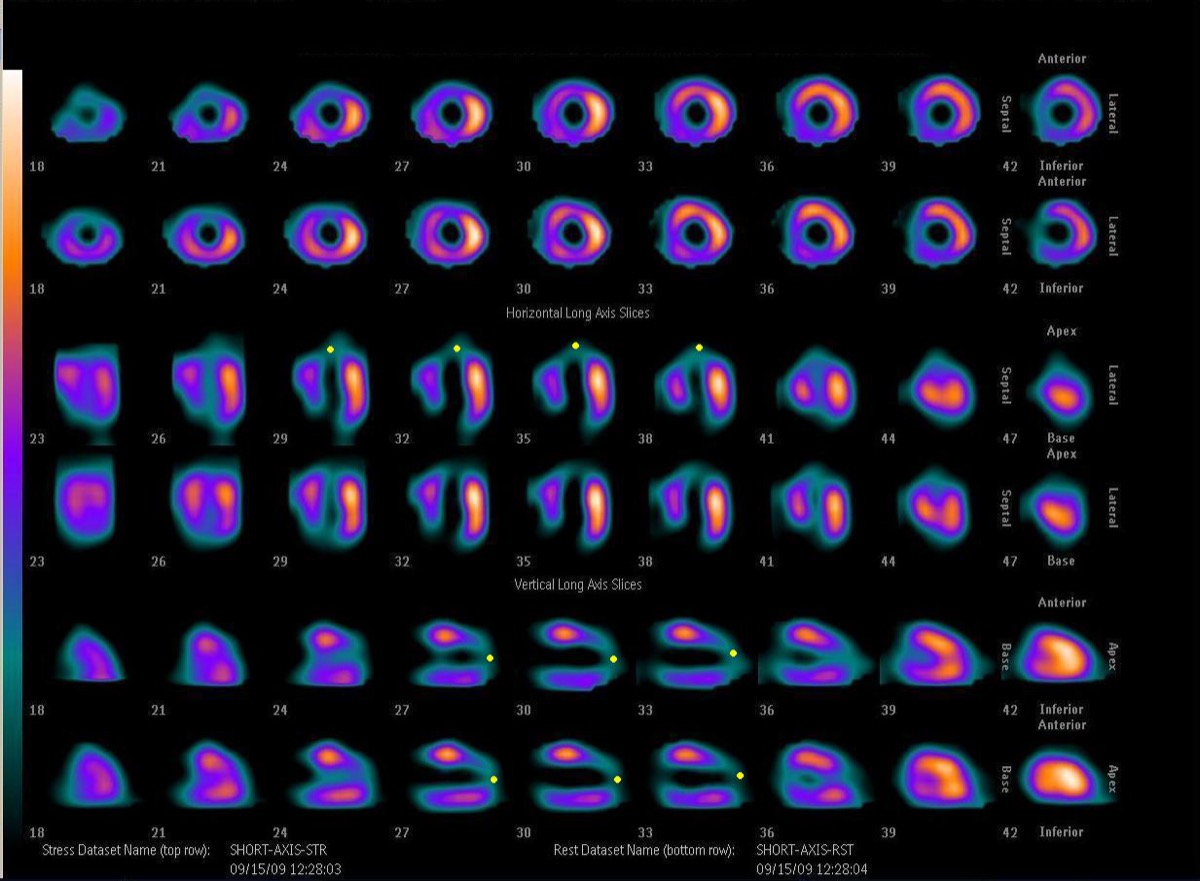 PET vs SPECT MPI: Absolute Myocardial Blood Flow
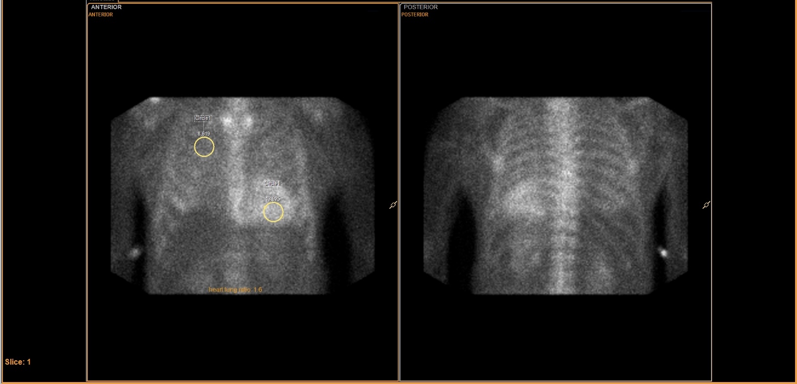 Tc-99m PYP Heart-to-Lung Ratio for Cardiac Amyloidosis
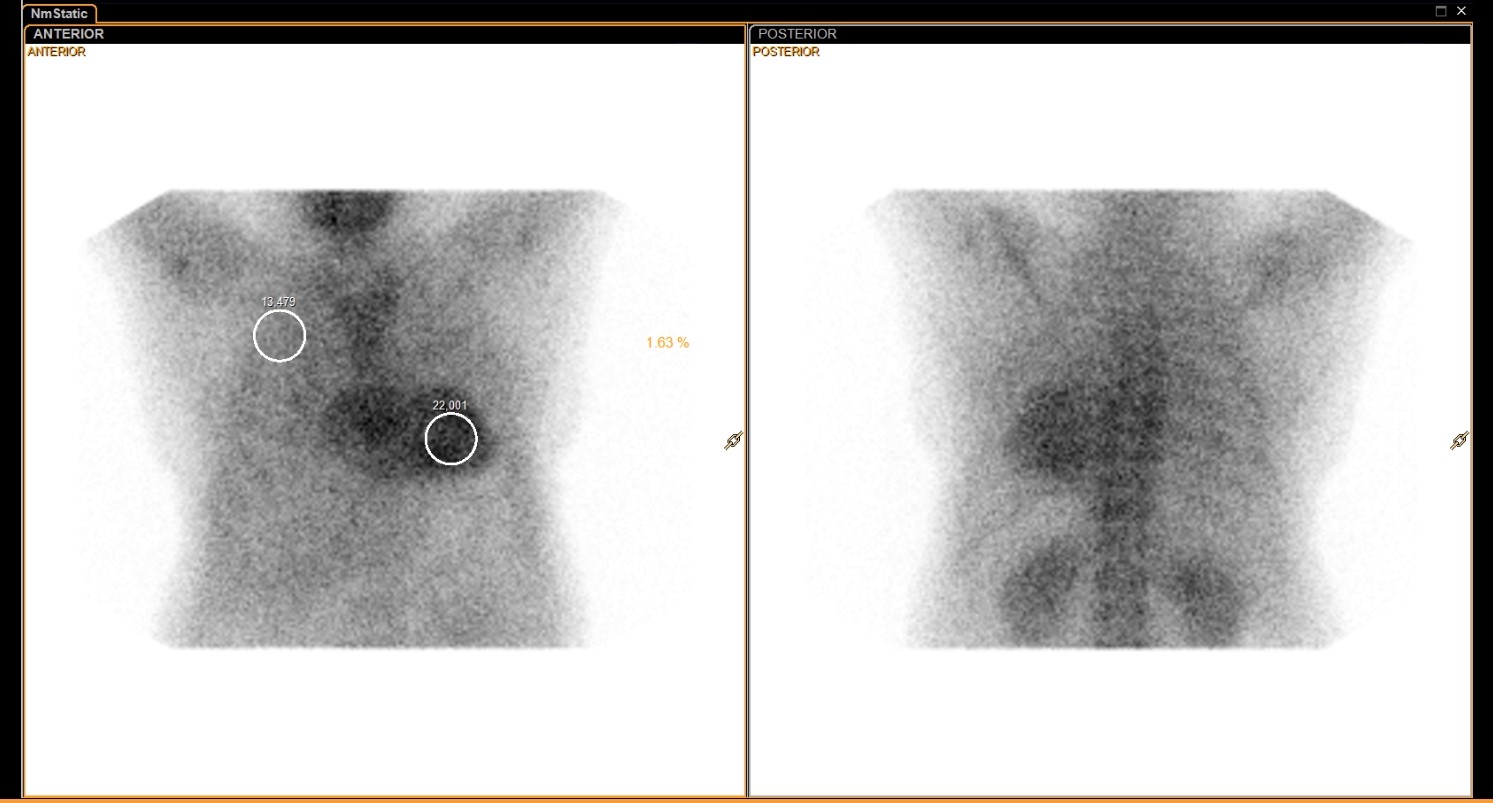 Tc-99m PYP Amyloid Scan Protocol
Normal Myocardial Perfusion SPECT, Stress and Rest
All 130 nuclear medicine images by category →
Practise image interpretation in the app
All 130 images as an interactive quiz, alongside 500 NMTCB and ARRT(N) questions with worked explanations, case studies and timed mock exams. 25 questions are free, no card and no account.