Tc-99m PYP Heart-to-Lung Ratio for Cardiac Amyloidosis
Provided clinical image
Question
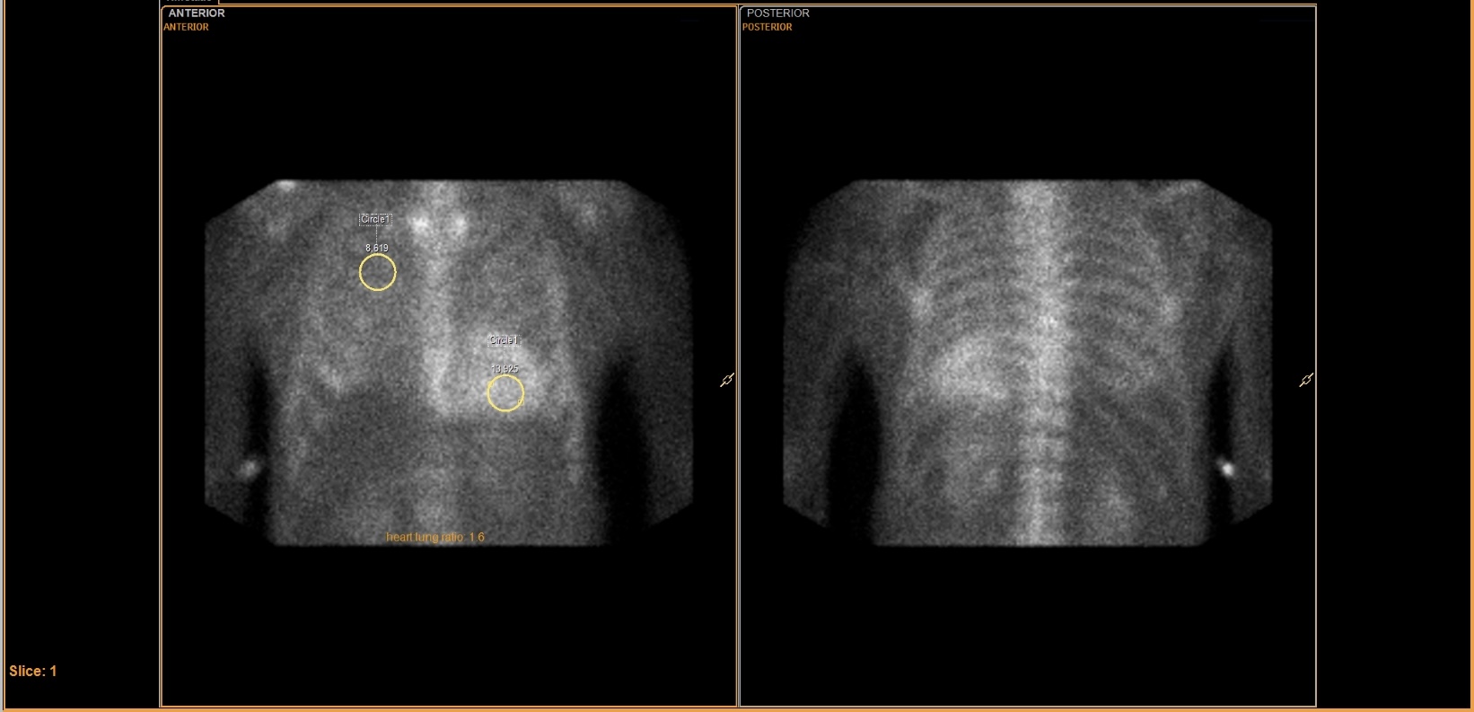
On this Tc-99m PYP scan, two ROI circles are drawn over the heart and lung, and the calculated heart-to-lung ratio is displayed as 1.6. What does this ratio indicate about the study result?
- AA heart-to-lung ratio ≥ 1.5 is considered positive for cardiac ATTR amyloidosis, and this value of 1.6 supports a positive result consistent with transthyretin amyloid cardiomyopathy
- BA heart-to-lung ratio of 1.6 is within the normal range for Tc-99m PYP studies, as ratios must exceed 2.0 before the scan is considered positive for cardiac amyloid deposition
- CThe heart-to-lung ratio is calculated only to confirm adequate tracer injection and does not contribute to the interpretation of cardiac amyloidosis on planar images
- DA heart-to-lung ratio ≥ 1.5 is a nonspecific finding seen in all bone-seeking radiopharmaceutical studies and cannot differentiate cardiac amyloid from other causes of cardiac uptake
Show the answer and explanation
A. A heart-to-lung ratio ≥ 1.5 is considered positive for cardiac ATTR amyloidosis, and this value of 1.6 supports a positive result consistent with transthyretin amyloid cardiomyopathy
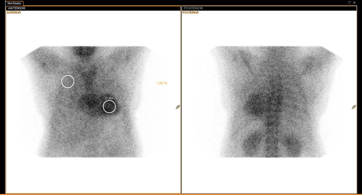
On Tc-99m pyrophosphate (PYP) cardiac amyloid imaging, a heart-to-contralateral lung (H/CL) ratio ≥ 1.5 measured at 1 hour post-injection is considered a positive result for ATTR (transthyretin) cardiac amyloidosis. A ratio of 1.6 exceeds this threshold and, when combined with visual grading, supports a positive study. This scan is used specifically to detect ATTR amyloid cardiomyopathy (both wild-type and hereditary forms) and is typically negative in AL (light-chain) amyloidosis.
More cardiac imaging images
 Tc-99m PYP Amyloid Scan Protocol
 Normal Myocardial Perfusion SPECT, Stress and Rest
 Myocardial Perfusion Tracers: Sestamibi and Tetrofosmin
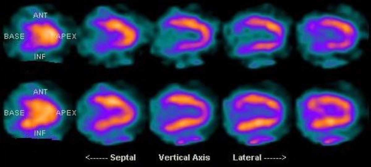 Diaphragmatic Attenuation Artifact on Cardiac SPECT
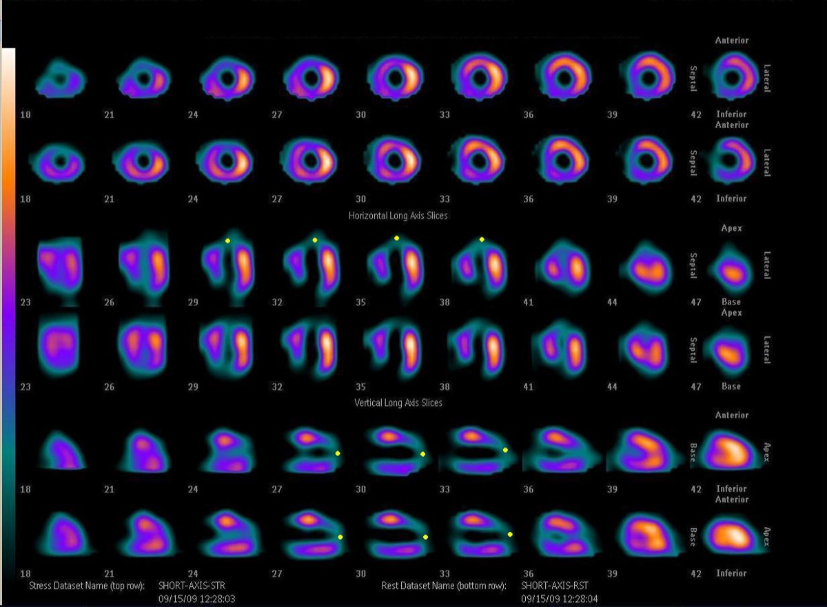 Fixed Perfusion Defect: Myocardial Scar on SPECT
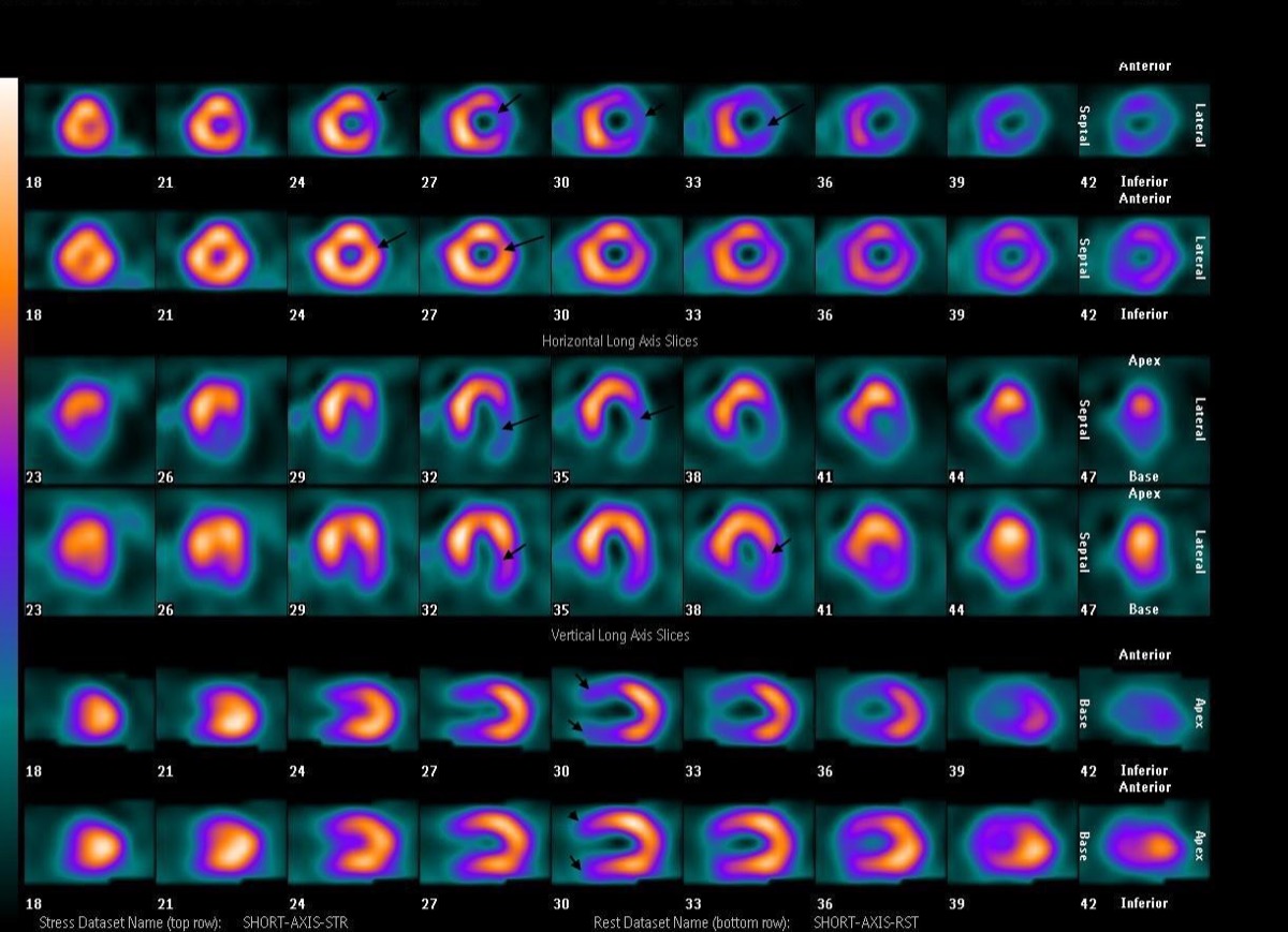 Reversible Lateral Wall Defect: Circumflex Ischemia
All 130 nuclear medicine images by category →
Practise image interpretation in the app
All 130 images as an interactive quiz, alongside 500 NMTCB and ARRT(N) questions with worked explanations, case studies and timed mock exams. 25 questions are free, no card and no account.