PET vs SPECT MPI: Absolute Myocardial Blood Flow
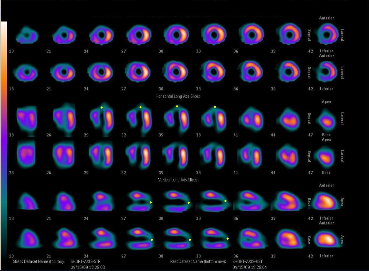
CC BY-SA 3.0 · Wikimedia Commons · source file
Question
PET myocardial perfusion imaging (with Rb-82 or N-13 ammonia) has several advantages over SPECT MPI. Which of the following is a key advantage of PET over SPECT for myocardial perfusion imaging?
- APET requires no attenuation correction
- BPET cannot detect multivessel disease
- CPET MPI can measure absolute myocardial blood flow (in mL/min/g) in addition to relative perfusion
- DPET has lower spatial resolution than SPECT
Show the answer and explanation
C. PET MPI can measure absolute myocardial blood flow (in mL/min/g) in addition to relative perfusion
A major advantage of PET MPI over SPECT is the ability to measure ABSOLUTE myocardial blood flow (MBF) in mL/min/g and myocardial flow reserve (MFR = stress MBF / rest MBF), using dynamic PET acquisitions with kinetic modeling. This is particularly valuable for detecting balanced multivessel ischemia (where all territories are equally ischemic, making relative perfusion appear normal) and microvascular dysfunction. PET also offers: superior spatial resolution, higher count rates, routine CT attenuation correction, and shorter acquisition times. Absolute MBF measurement is not possible with conventional SPECT.
More cardiac imaging images
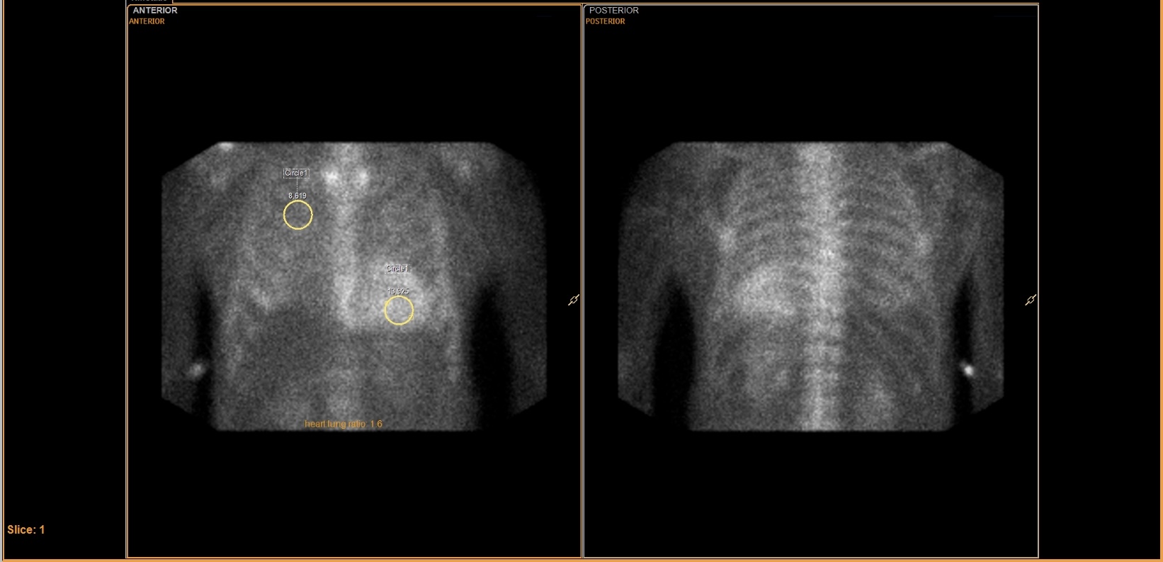 Tc-99m PYP Heart-to-Lung Ratio for Cardiac Amyloidosis
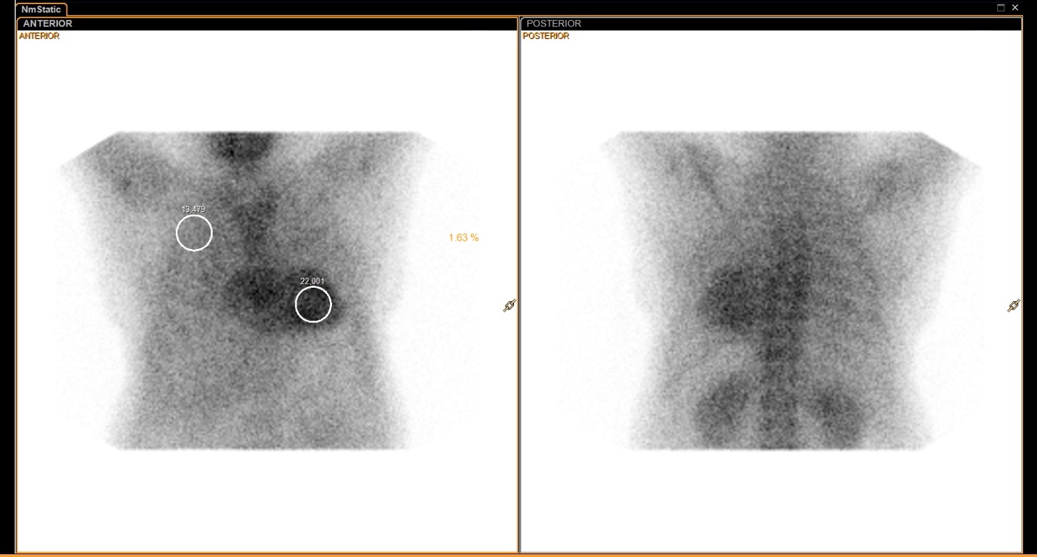 Tc-99m PYP Amyloid Scan Protocol
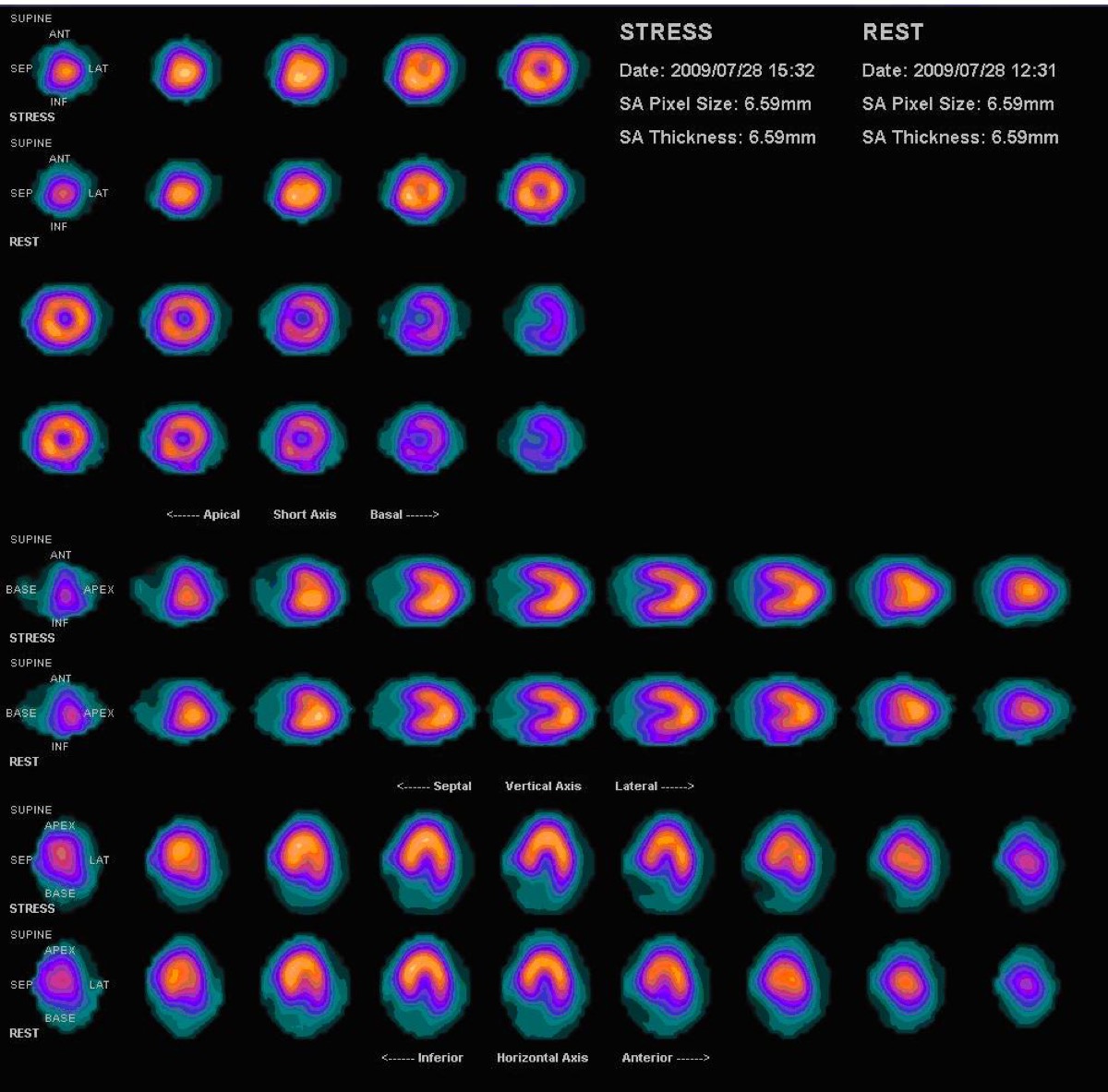 Normal Myocardial Perfusion SPECT, Stress and Rest
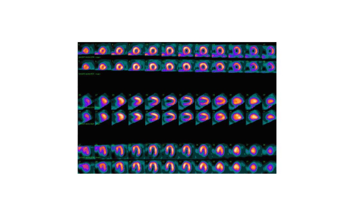 Myocardial Perfusion Tracers: Sestamibi and Tetrofosmin
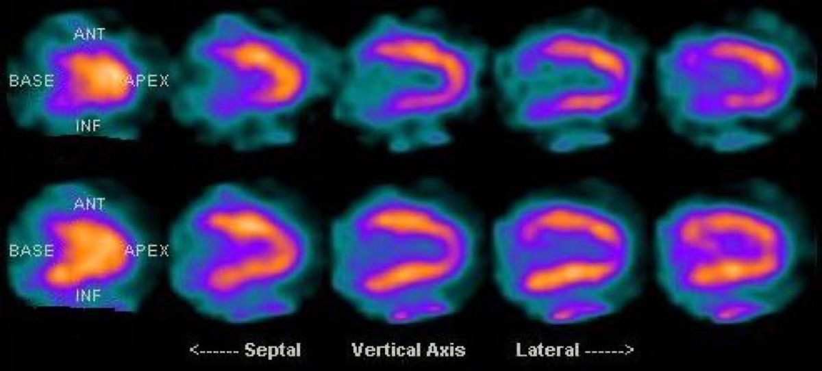 Diaphragmatic Attenuation Artifact on Cardiac SPECT
Fixed Perfusion Defect: Myocardial Scar on SPECT
All 130 nuclear medicine images by category →
Practise image interpretation in the app
All 130 images as an interactive quiz, alongside 500 NMTCB and ARRT(N) questions with worked explanations, case studies and timed mock exams. 25 questions are free, no card and no account.