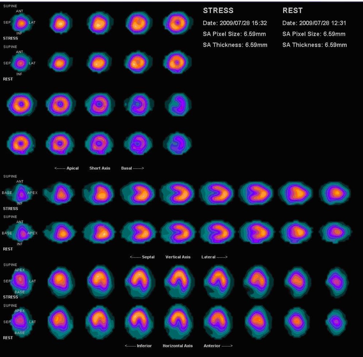
Prominent Right Ventricle on MPI: RV Hypertrophy
CC BY 3.0 · Myohan / Wikimedia Commons · source file
Question
On a standard myocardial perfusion SPECT study, the right ventricle (RV) is normally faintly visible or not seen at all. Prominent, easily visible RV uptake on MPI is a significant finding because it suggests:
- ANormal variant in all patients
- BArtifact from diaphragmatic attenuation
- CRV ischemia from right coronary artery disease
- DRight ventricular hypertrophy (RVH) or elevated RV pressure (pulmonary hypertension, severe tricuspid regurgitation)
Show the answer and explanation
D. Right ventricular hypertrophy (RVH) or elevated RV pressure (pulmonary hypertension, severe tricuspid regurgitation)
The right ventricle is normally thin-walled (much thinner than the LV), so it takes up proportionally less tracer and is usually faintly seen or invisible on standard dose MPI. When the RV myocardium is significantly hypertrophied (from chronically elevated RV pressure — as in pulmonary arterial hypertension, cor pulmonale, or significant RV outflow obstruction), its mass is increased and it becomes prominently visible on MPI, similar in intensity to the LV. Prominent RV visualization is therefore a marker for RV pressure overload. This finding can also be seen in RV infarction (from RCA occlusion proximal to the RV branches), where it may appear as an RV perfusion defect.
More cardiac imaging images
 Dobutamine Stress for Myocardial Perfusion Imaging
 PET vs SPECT MPI: Absolute Myocardial Blood Flow
 Tc-99m PYP Heart-to-Lung Ratio for Cardiac Amyloidosis
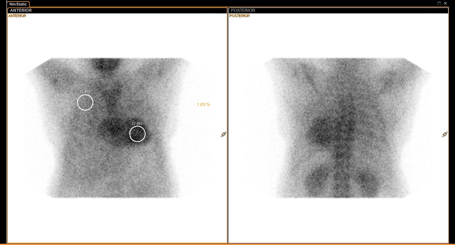 Tc-99m PYP Amyloid Scan Protocol
Normal Myocardial Perfusion SPECT, Stress and Rest
Myocardial Perfusion Tracers: Sestamibi and Tetrofosmin
All 130 nuclear medicine images by category →
Practise image interpretation in the app
All 130 images as an interactive quiz, alongside 500 NMTCB and ARRT(N) questions with worked explanations, case studies and timed mock exams. 25 questions are free, no card and no account.