Multivessel Coronary Disease on Stress MPI
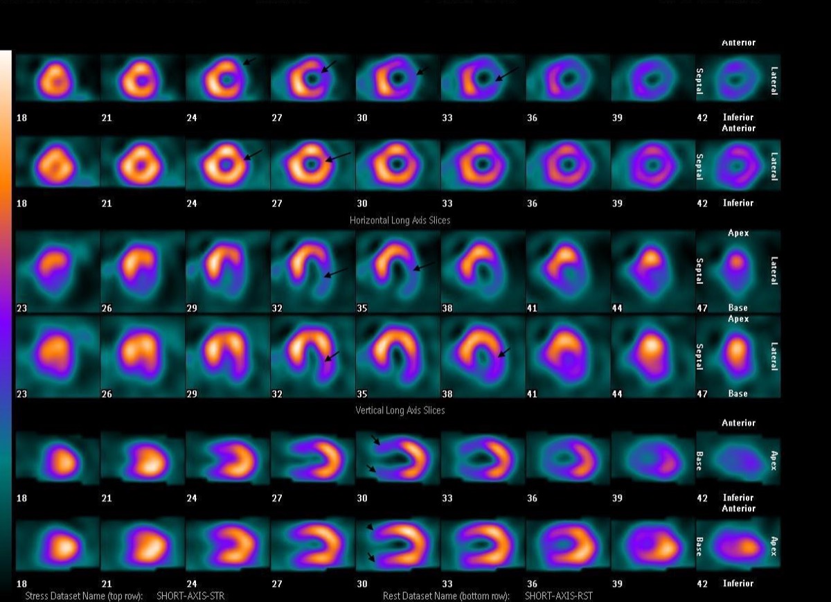
CC BY-SA 4.0 · Wikimedia Commons · source file
Question
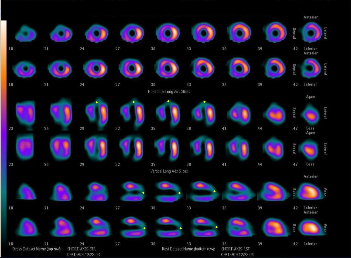
A patient has reversible perfusion defects in BOTH the anterior wall AND the inferior wall on stress MPI, with an LVEF drop from 60% to 45% with stress. Which of the following is the most appropriate interpretation?
- ATwo separate prior myocardial infarctions (fixed defects)
- BNormal variant — bilateral attenuation artifacts
- CBalanced triple-vessel ischemia — stress perfusion appears normal
- DMultivessel CAD — ischemia in both LAD and RCA territories with stress-induced LV dysfunction
Show the answer and explanation
D. Multivessel CAD — ischemia in both LAD and RCA territories with stress-induced LV dysfunction
Reversible perfusion defects in multiple coronary territories (anterior wall = LAD, inferior wall = RCA) combined with a significant LVEF drop at stress (>5%) indicates multivessel CAD with hemodynamically significant ischemia in two major territories. The stress-induced LV dysfunction (transient ischemic dilation) indicates that the ischemic burden is large enough to impair global left ventricular function. This high-risk scan pattern warrants urgent cardiology consultation and likely coronary angiography. Multivessel disease with LV dysfunction at stress carries a high annual cardiac event rate (>3%) and typically benefits from revascularization.
More cardiac imaging images
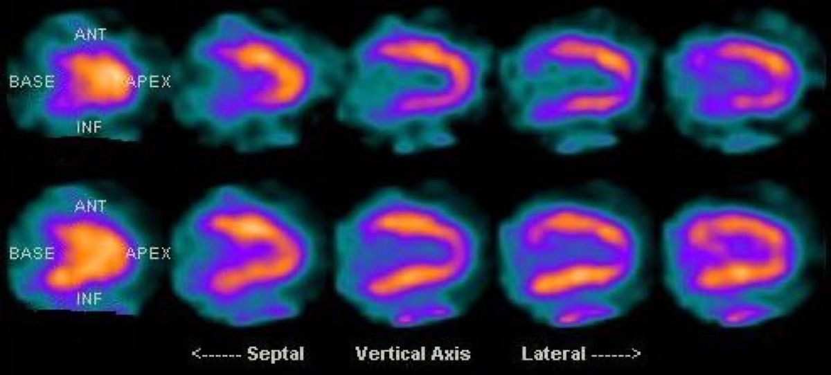 Prone Imaging in Cardiac SPECT
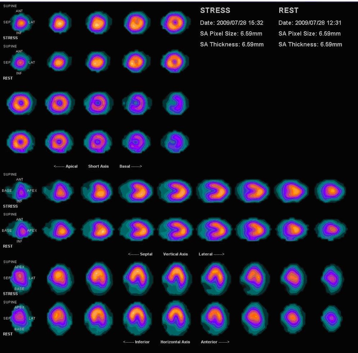 Prominent Right Ventricle on MPI: RV Hypertrophy
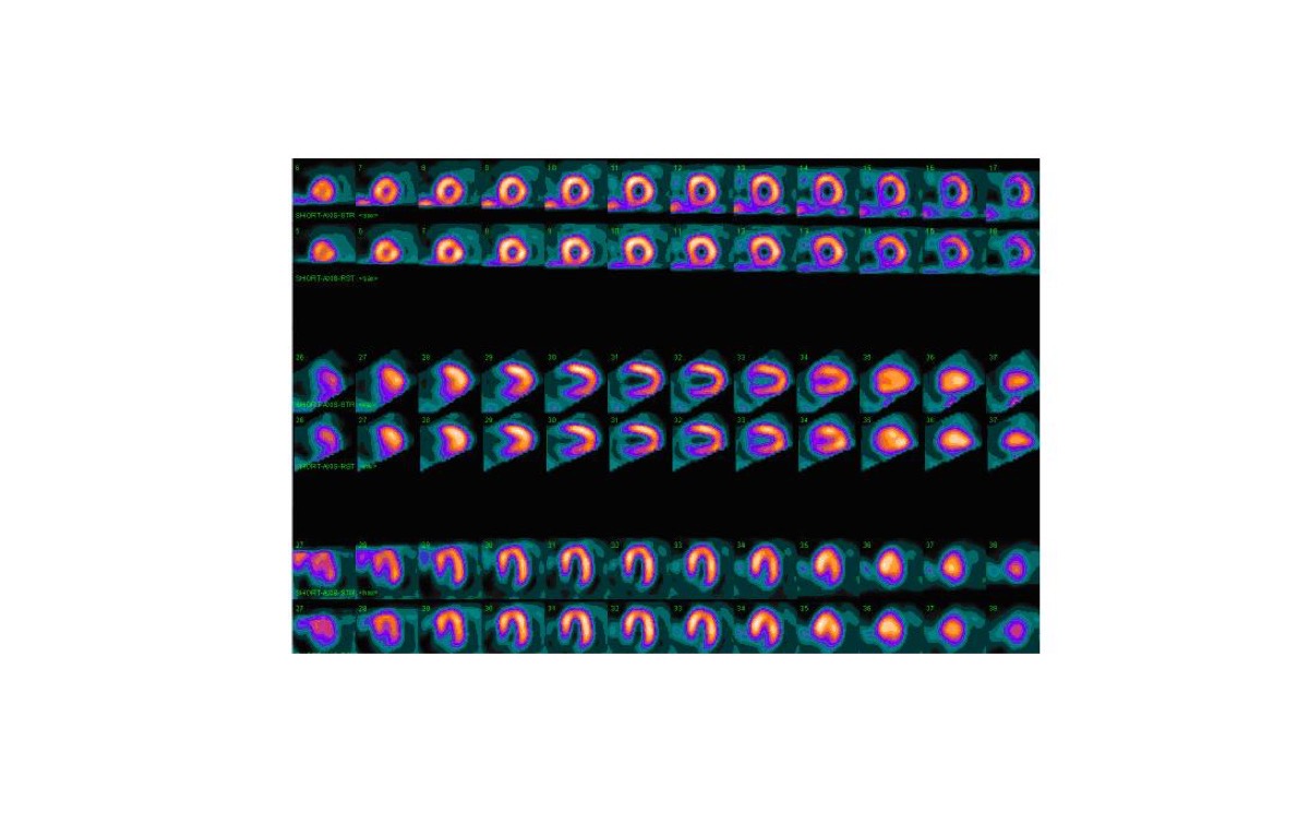 Dobutamine Stress for Myocardial Perfusion Imaging
 PET vs SPECT MPI: Absolute Myocardial Blood Flow
 Tc-99m PYP Heart-to-Lung Ratio for Cardiac Amyloidosis
 Tc-99m PYP Amyloid Scan Protocol
All 130 nuclear medicine images by category →
Practise image interpretation in the app
All 130 images as an interactive quiz, alongside 500 NMTCB and ARRT(N) questions with worked explanations, case studies and timed mock exams. 25 questions are free, no card and no account.