Tc-99m PYP Amyloid Scan Protocol
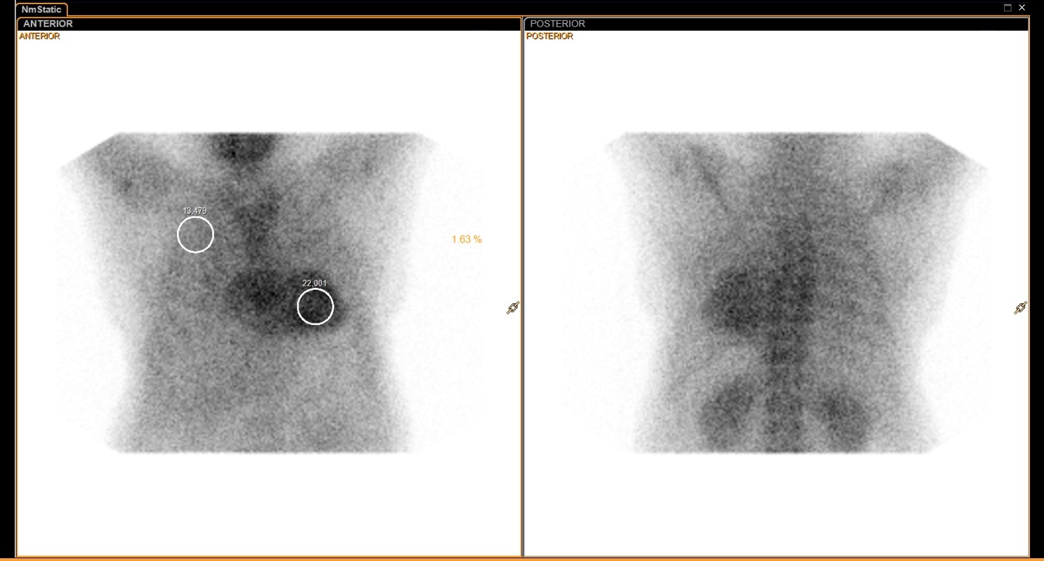
Provided clinical image
Question
On this Tc-99m PYP amyloid scan, planar chest images are acquired with ROI circles placed over the heart and lung. According to standard protocol, at what timepoint are these planar images obtained after injection, and why?
- APlanar images are acquired immediately after injection (0–5 minutes) to capture peak cardiac blood-pool activity before the tracer redistributes into bone and soft tissue
- BPlanar images for the heart-to-lung ratio are acquired at 1 hour post-injection, allowing adequate blood-pool clearance while capturing tracer uptake in amyloid deposits for quantification
- CPlanar images are obtained at 48–72 hours post-injection, similar to Ga-67 imaging, because Tc-99m PYP requires an extended delay for amyloid-specific uptake to reach detectable levels
- DPlanar images are acquired only after SPECT confirms cardiac uptake at 3 hours, since planar imaging alone cannot generate a valid heart-to-lung ratio without prior tomographic confirmation
Show the answer and explanation
D. Planar images are acquired only after SPECT confirms cardiac uptake at 3 hours, since planar imaging alone cannot generate a valid heart-to-lung ratio without prior tomographic confirmation
Standard Tc-99m PYP cardiac amyloid protocol includes SPECT imaging at 3 hours post-injection to confirm true myocardial uptake and differentiate it from residual blood pool activity. Planar anterior and posterior chest images for the heart-to-contralateral lung (H/CL) ratio are obtained in conjunction with SPECT. An H/CL ratio ≥ 1.5, combined with SPECT confirmation of myocardial uptake, supports a positive result for ATTR cardiac amyloidosis.
More cardiac imaging images
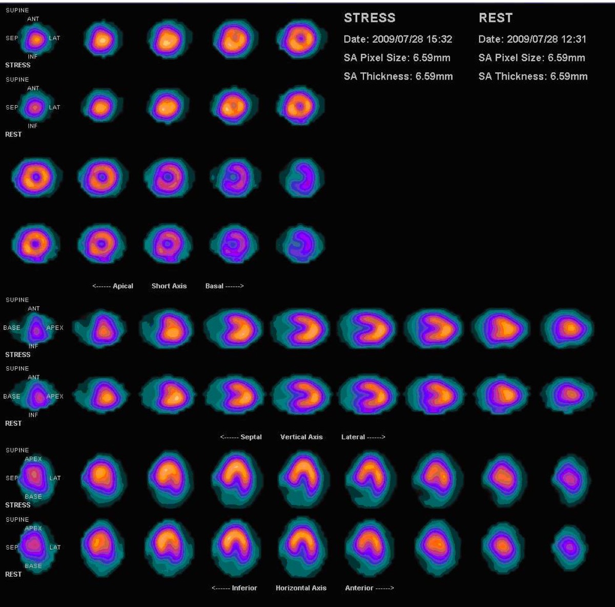 Normal Myocardial Perfusion SPECT, Stress and Rest
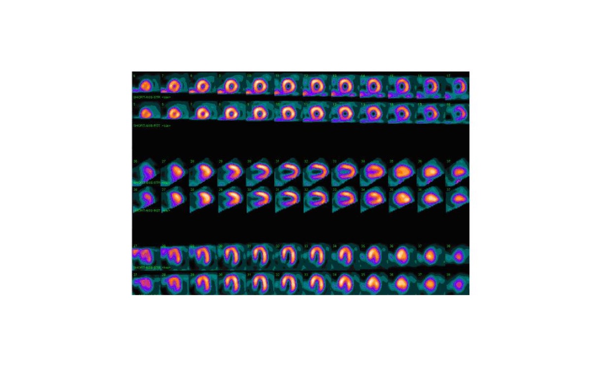 Myocardial Perfusion Tracers: Sestamibi and Tetrofosmin
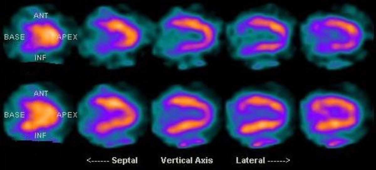 Diaphragmatic Attenuation Artifact on Cardiac SPECT
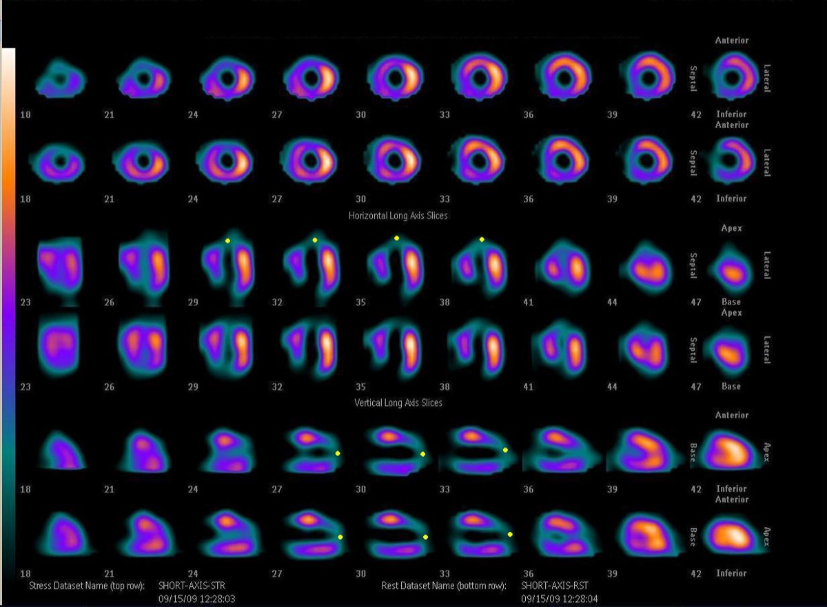 Fixed Perfusion Defect: Myocardial Scar on SPECT
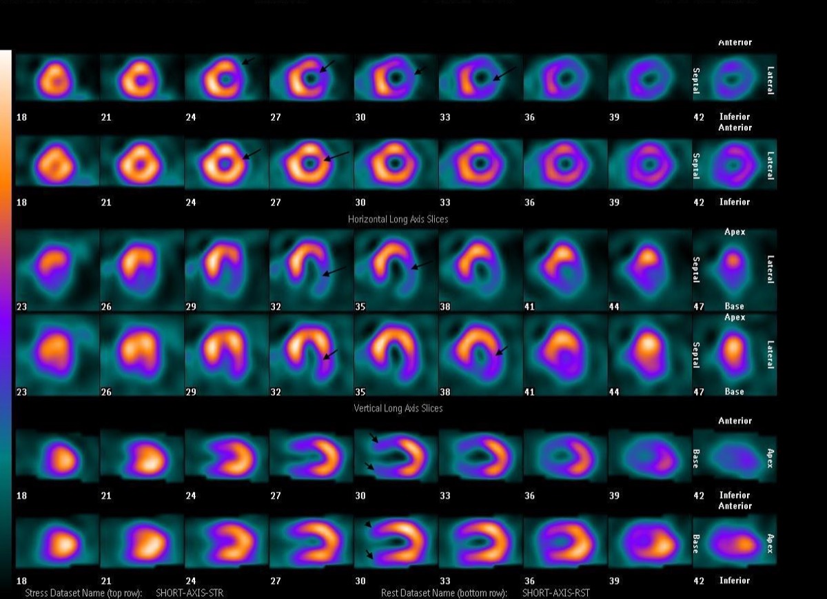 Reversible Lateral Wall Defect: Circumflex Ischemia
Bull’s-Eye Polar Map: What the Center Shows
All 130 nuclear medicine images by category →
Practise image interpretation in the app
All 130 images as an interactive quiz, alongside 500 NMTCB and ARRT(N) questions with worked explanations, case studies and timed mock exams. 25 questions are free, no card and no account.