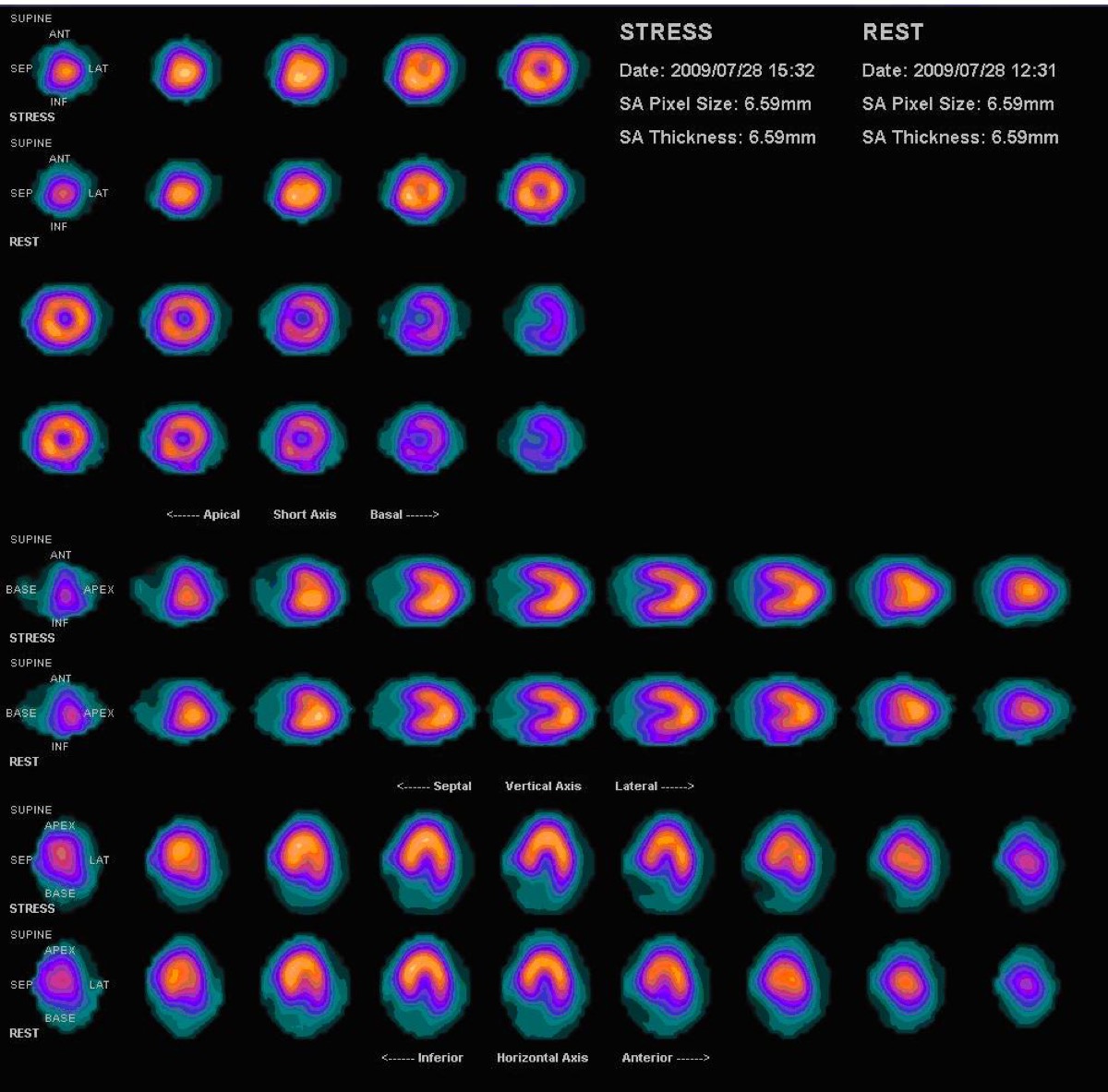
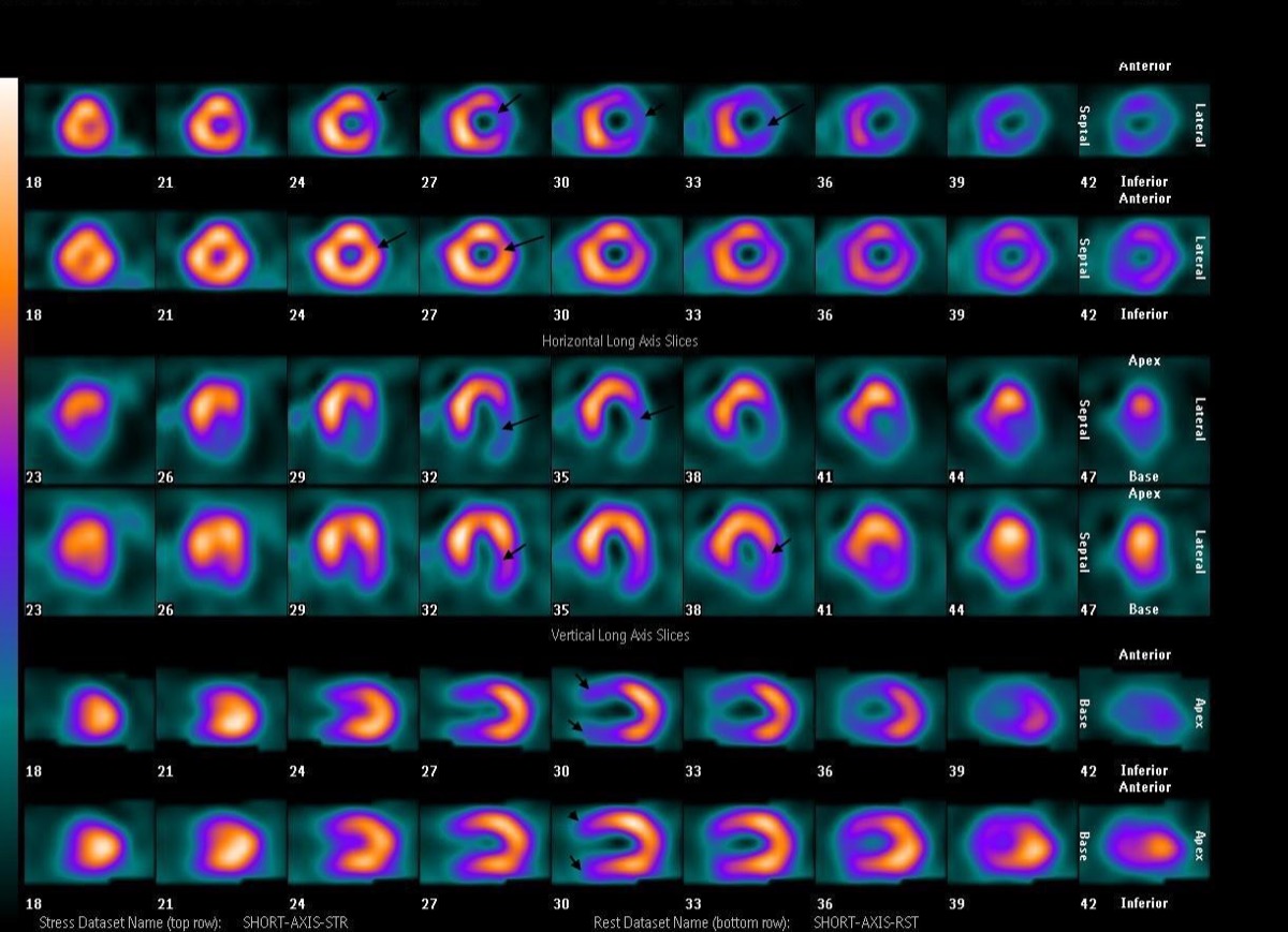
Normal Myocardial Perfusion SPECT, Stress and Rest
CC BY 3.0 · Myohan / Wikimedia Commons · source file
Question
This myocardial perfusion SPECT shows uniform tracer uptake in all myocardial segments on both stress and rest images, with no perfusion defects. What is the most appropriate interpretation?
- ANormal study — no evidence of significant CAD
- BBalanced ischemia in all three coronary territories
- CFixed anterior wall defect consistent with prior MI
- DInferior wall attenuation artifact
Show the answer and explanation
A. Normal study — no evidence of significant CAD
A normal myocardial perfusion SPECT shows homogeneous, uniform uptake throughout the left ventricular myocardium at both stress and rest, with no areas of relative decreased uptake. This indicates adequate perfusion to all coronary territories and makes significant obstructive CAD unlikely. Balanced ischemia is a rare cause of a normal-appearing scan (all territories equally ischemic), but uniform normal uptake throughout all segments is most consistent with a truly normal study.
More cardiac imaging images
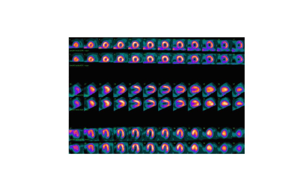 Myocardial Perfusion Tracers: Sestamibi and Tetrofosmin
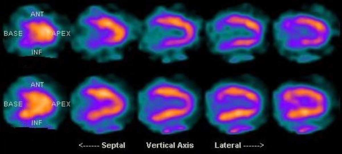 Diaphragmatic Attenuation Artifact on Cardiac SPECT
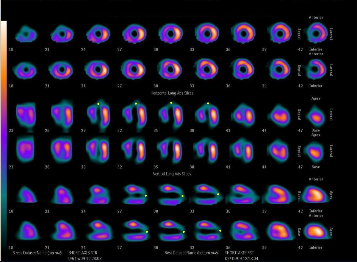 Fixed Perfusion Defect: Myocardial Scar on SPECT
 Reversible Lateral Wall Defect: Circumflex Ischemia
Bull’s-Eye Polar Map: What the Center Shows
Anteroapical Fixed Defect: LAD Infarction on MPI
All 130 nuclear medicine images by category →
Practise image interpretation in the app
All 130 images as an interactive quiz, alongside 500 NMTCB and ARRT(N) questions with worked explanations, case studies and timed mock exams. 25 questions are free, no card and no account.