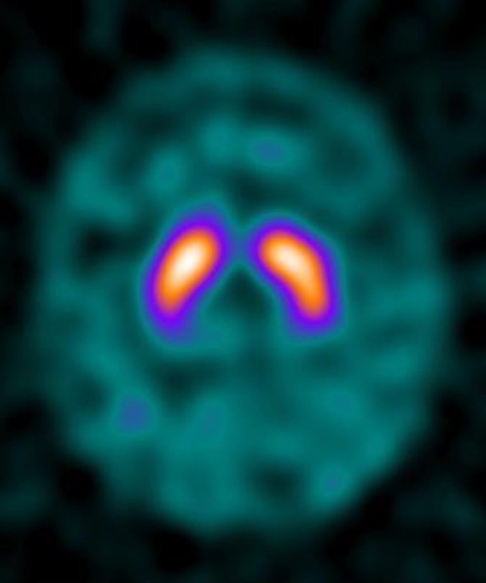
Parkinson’s Disease on DaTscan: Loss of the Comma Shape
Public domain · Wikimedia Commons · source file
Question
In a patient with Parkinson's disease, a DaTSCAN (I-123 ioflupane SPECT) would show which characteristic pattern?
- ANormal bilateral comma-shaped striatal uptake
- BBilateral increased striatal uptake
- CAbsent uptake in the caudate only, with preserved putamen
- DAsymmetric reduction of putamen uptake — loss of the comma shape to a period/dot shape
Show the answer and explanation
D. Asymmetric reduction of putamen uptake — loss of the comma shape to a period/dot shape
In Parkinson's disease, the dopaminergic neurons projecting from the substantia nigra to the putamen degenerate preferentially. On DaTSCAN, this produces asymmetric (contralateral to clinically affected side) reduction in putamen uptake, converting the normal 'comma' shape to a 'period' or 'egg' as the posterior putamen loses dopamine transporter density. More advanced PD shows loss of both putamen and caudate uptake bilaterally. Normal DaTSCAN (bilateral full commas) excludes primary Parkinson's disease. Drug-induced parkinsonism typically shows a normal DaTSCAN.
More brain imaging images
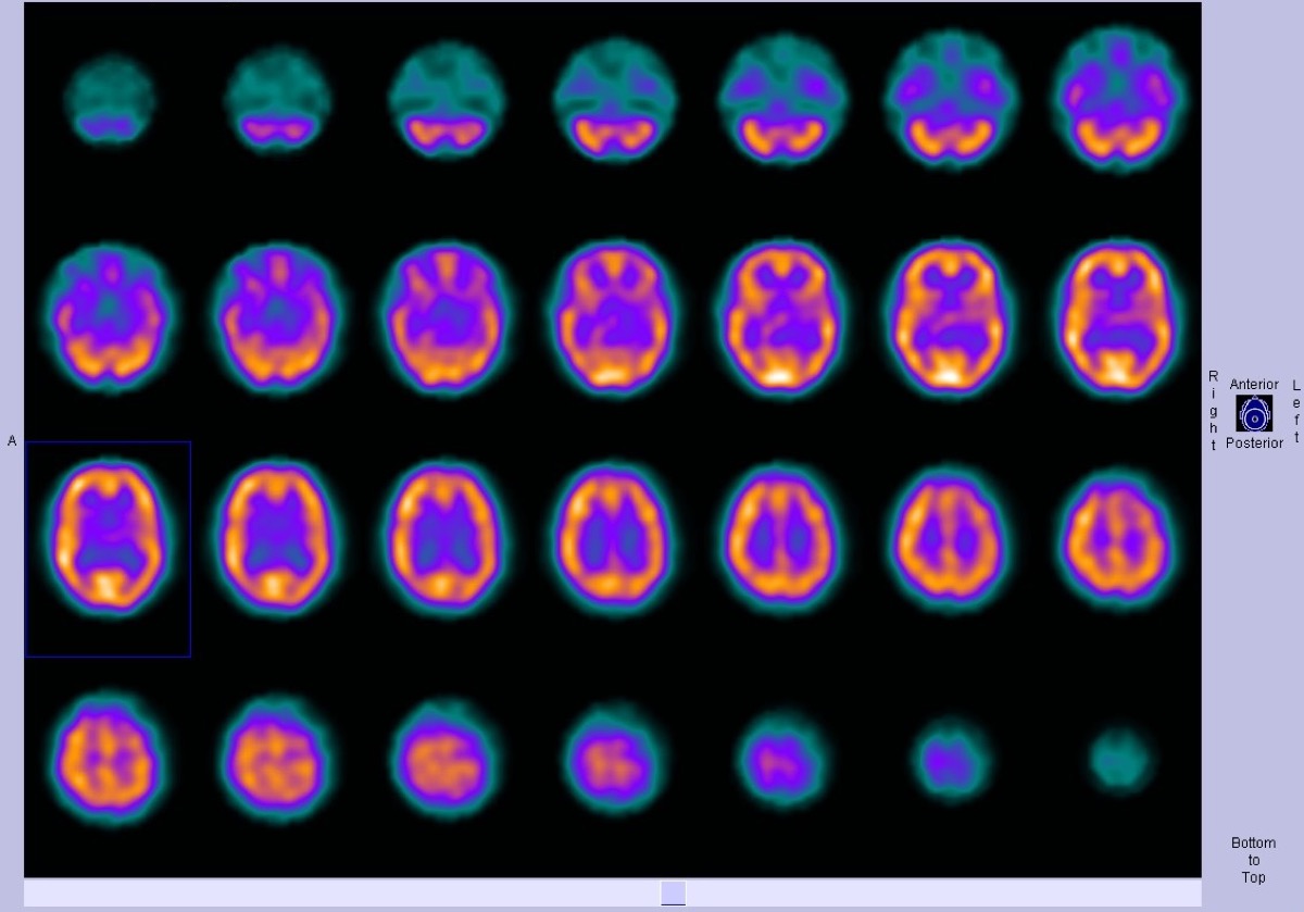 Frontotemporal Dementia on Brain Perfusion SPECT
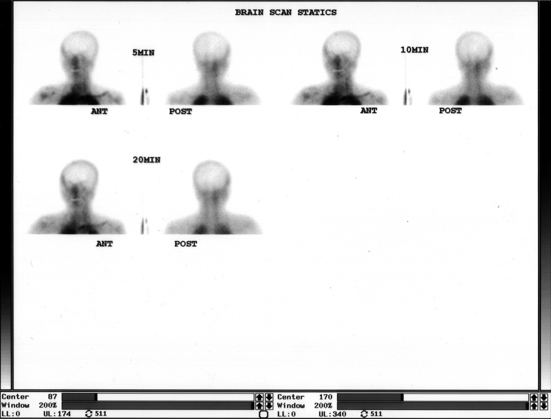 Brain Death Scan: Absent Cerebral Perfusion
Ictal SPECT: Hyperperfusion at the Seizure Focus
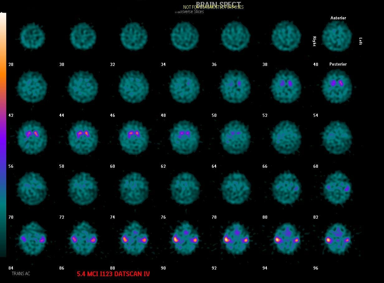 Dementia with Lewy Bodies on DaTscan
Alzheimer’s Disease on FDG PET
 Focal Perfusion Defect on Brain SPECT
All 130 nuclear medicine images by category →
Practise image interpretation in the app
All 130 images as an interactive quiz, alongside 500 NMTCB and ARRT(N) questions with worked explanations, case studies and timed mock exams. 25 questions are free, no card and no account.