Brain Death Scan: Absent Cerebral Perfusion
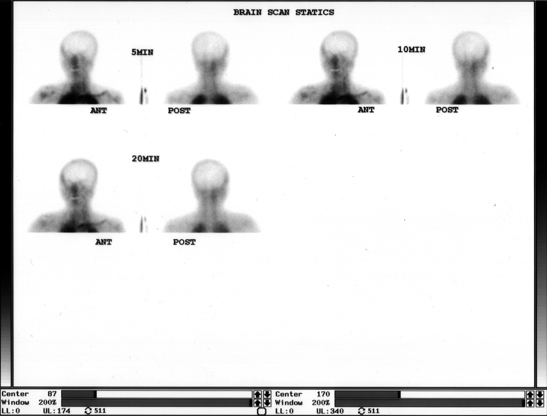
Provided clinical image
Question
A brain death radionuclide cerebral blood flow study is performed in a comatose patient on a ventilator. The study shows complete absence of cerebral perfusion (no activity in the cerebral hemispheres or cerebellum) with visualization only of scalp and skull activity ('hollow skull sign'). This finding is diagnostic of:
- ASevere head injury with residual perfusion
- BLocked-in syndrome
- CPersistent vegetative state
- DBrain death — no cerebral blood flow confirming cessation of all brain function
Show the answer and explanation
D. Brain death — no cerebral blood flow confirming cessation of all brain function
Radionuclide cerebral blood flow imaging (using Tc-99m HMPAO or Tc-99m DTPA) can confirm brain death by demonstrating complete absence of cerebral perfusion. In brain death, the markedly elevated intracranial pressure (equal to or exceeding systemic perfusion pressure) prevents blood from entering the cerebral circulation. The study shows: (1) absent intracranial activity; (2) preserved scalp and skull blood flow ('hollow skull' or 'empty light bulb' sign); (3) absent posterior fossa/cerebellar perfusion. This nuclear medicine finding is one of several ancillary confirmatory tests for brain death when clinical assessment alone is insufficient (e.g., pharmacologic sedation, metabolic disturbances).
More brain imaging images
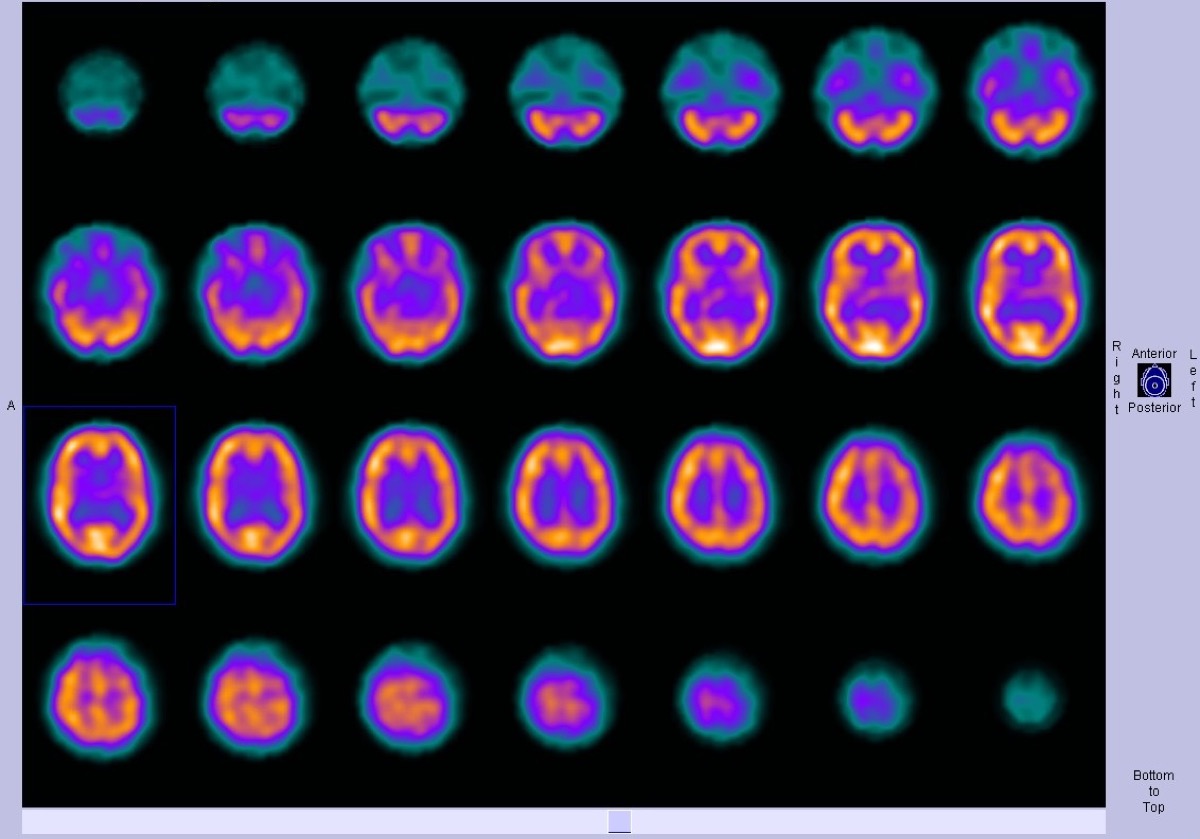 Ictal SPECT: Hyperperfusion at the Seizure Focus
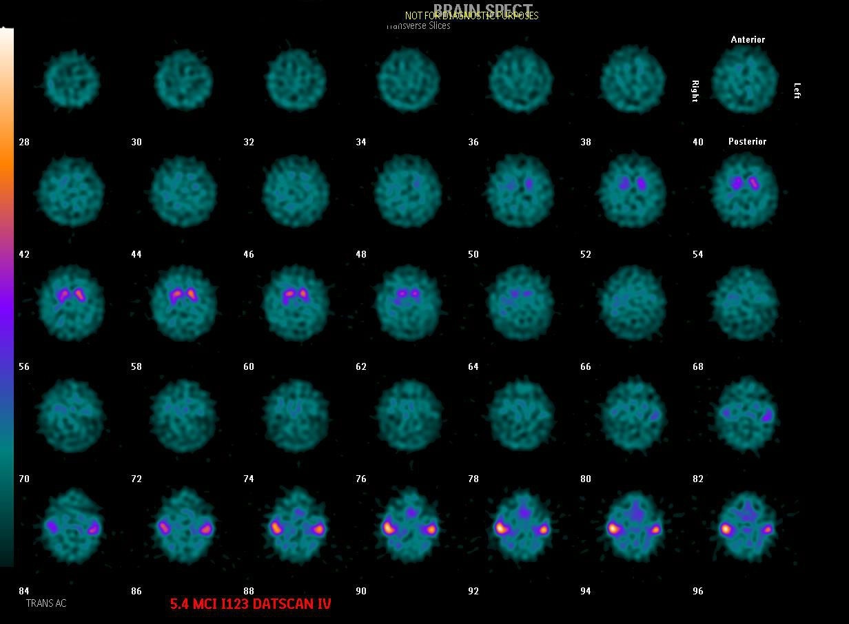 Dementia with Lewy Bodies on DaTscan
Alzheimer’s Disease on FDG PET
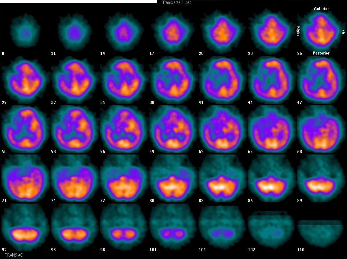 Focal Perfusion Defect on Brain SPECT
Brain Perfusion SPECT: Injection Environment
 Normal Tc-99m ECD Brain SPECT: Gray vs White Matter
All 130 nuclear medicine images by category →
Practise image interpretation in the app
All 130 images as an interactive quiz, alongside 500 NMTCB and ARRT(N) questions with worked explanations, case studies and timed mock exams. 25 questions are free, no card and no account.