Alzheimer’s Disease on Brain Perfusion SPECT
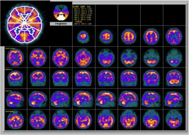
Provided clinical image
Question
Brain perfusion SPECT is used to evaluate regional cerebral blood flow. In a patient with dementia, Alzheimer's disease classically produces which perfusion pattern?
- AFrontal and anterior temporal hypoperfusion
- BUnilateral frontal and basal ganglia hypoperfusion
- CBilateral parietal and posterior temporal hypoperfusion
- DDiffuse symmetric cortical hypoperfusion
Show the answer and explanation
C. Bilateral parietal and posterior temporal hypoperfusion
Alzheimer's disease characteristically produces bilateral parietal and posterior temporal hypoperfusion (decreased uptake) on brain perfusion SPECT and FDG-PET. This reflects the pattern of early neurodegeneration in AD (posterior cortex, precuneus, posterior cingulate). Frontotemporal dementia (FTD) shows frontal and anterior temporal hypoperfusion. Unilateral basal ganglia abnormalities suggest vascular disease. Diffuse symmetric hypoperfusion may be seen in medication effects.
More brain imaging images
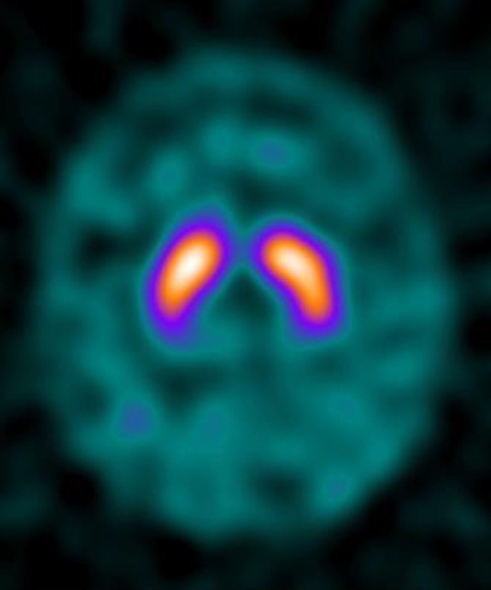 Parkinson’s Disease on DaTscan: Loss of the Comma Shape
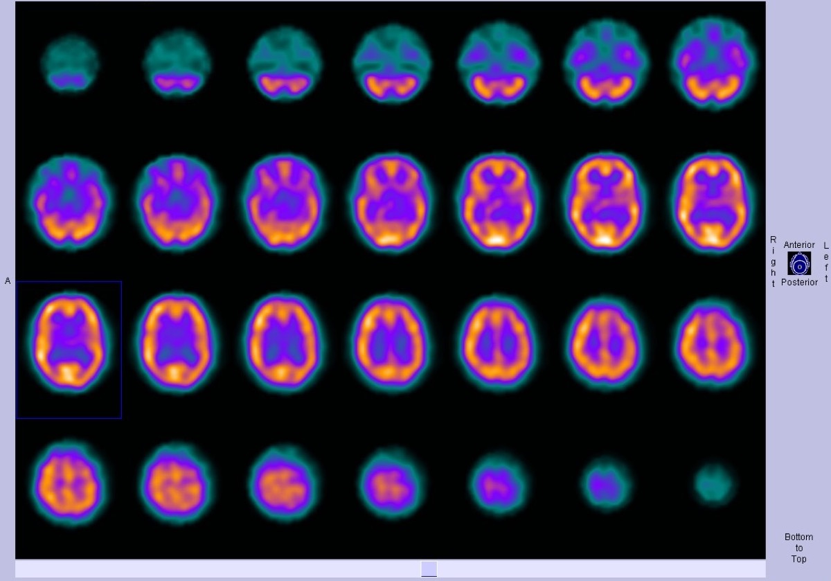 Frontotemporal Dementia on Brain Perfusion SPECT
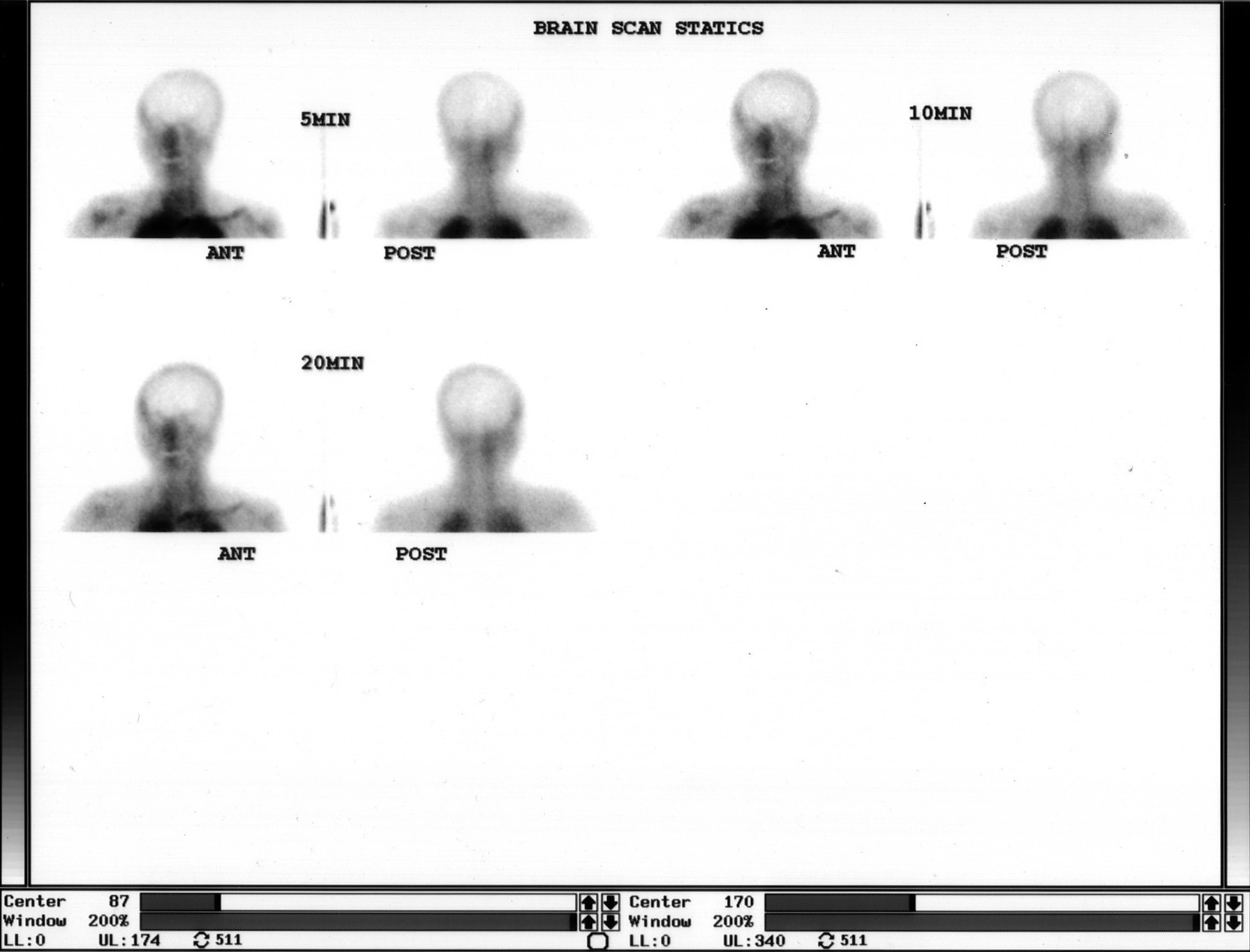 Brain Death Scan: Absent Cerebral Perfusion
Ictal SPECT: Hyperperfusion at the Seizure Focus
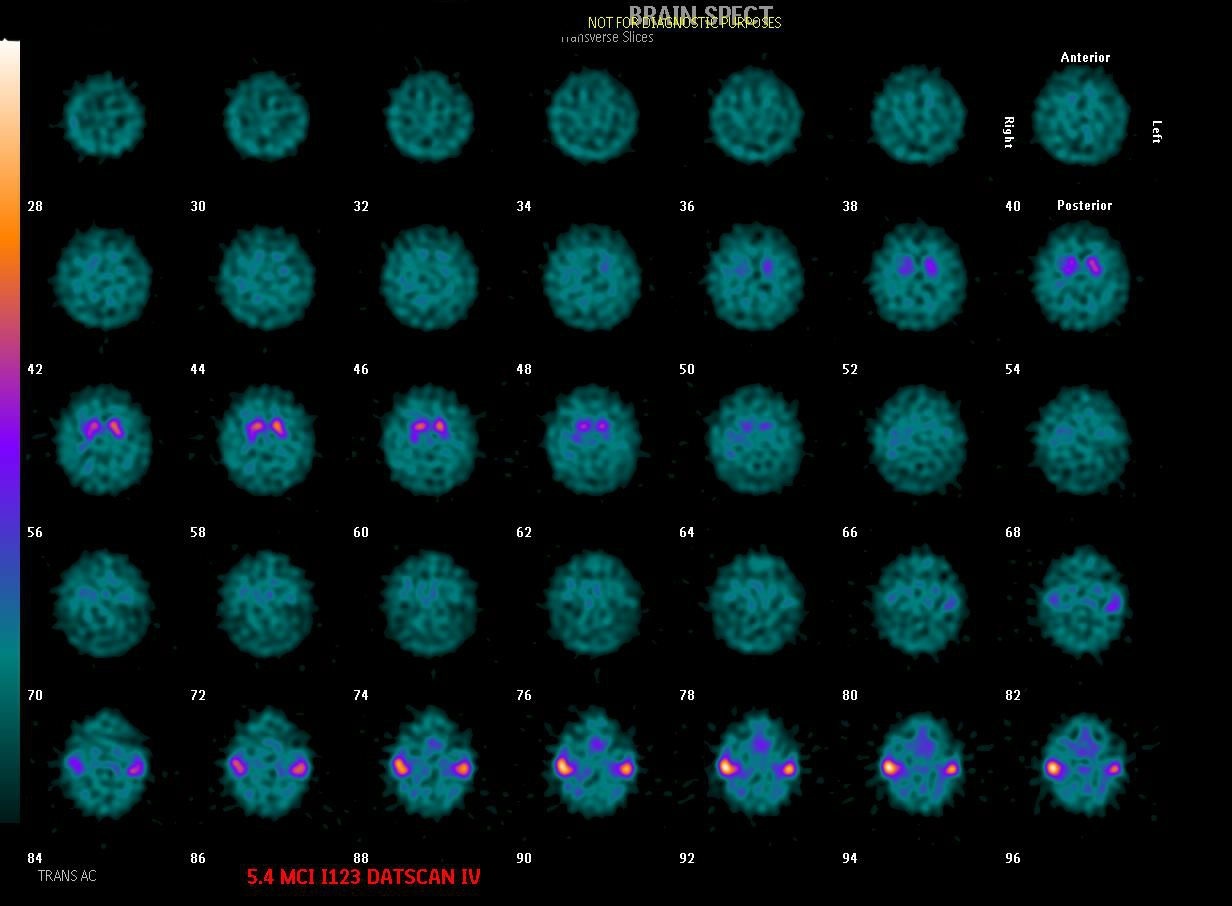 Dementia with Lewy Bodies on DaTscan
Alzheimer’s Disease on FDG PET
All 130 nuclear medicine images by category →
Practise image interpretation in the app
All 130 images as an interactive quiz, alongside 500 NMTCB and ARRT(N) questions with worked explanations, case studies and timed mock exams. 25 questions are free, no card and no account.