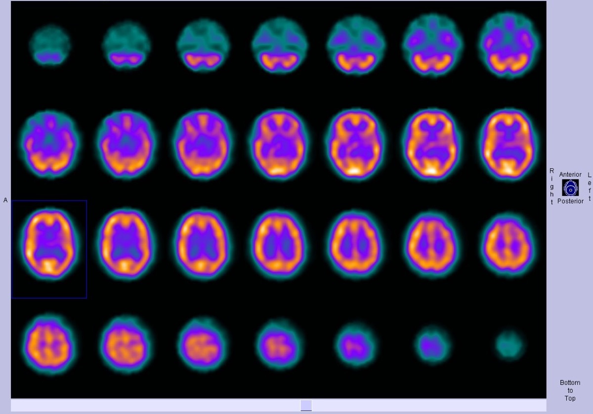
Frontotemporal Dementia on Brain Perfusion SPECT
CC BY-SA 3.0 · Dr Bruno Di Muzio / Radiopaedia / Wikimedia Commons · source file
Question
Brain perfusion SPECT in a patient with progressive personality changes, disinhibition, and executive dysfunction (but relatively preserved memory early in the disease) shows bilateral FRONTAL and ANTERIOR TEMPORAL hypoperfusion with relative sparing of parietal cortex. This pattern is most consistent with:
- AFrontotemporal dementia (FTD) — the classic anterior-predominant hypoperfusion pattern
- BAlzheimer's disease — typically causing bilateral posterior parietal and temporoparietal hypoperfusion with relative preservation of frontal and primary sensorimotor cortices until advanced stages
- CVascular dementia — characterized by asymmetric, multifocal cortical and subcortical hypoperfusion in a vascular territory distribution reflecting cumulative ischemic white matter injury
- DLewy body dementia — producing posterior occipital hypoperfusion with relative cingulate cortex sparing (the cingulate island sign), accompanied by reduced dopaminergic striatal tracer binding on DaTscan
Show the answer and explanation
A. Frontotemporal dementia (FTD) — the classic anterior-predominant hypoperfusion pattern
Frontotemporal dementia (FTD) produces bilateral frontal and anterior temporal hypoperfusion on brain perfusion SPECT and FDG-PET, reflecting the anterior-predominant neurodegeneration in this condition. This distinguishes it from Alzheimer's disease (posterior parietal and temporal hypoperfusion) and Lewy body dementia (parieto-occipital hypoperfusion, often with relative sparing of the posterior cingulate cortex). FTD clinical features — disinhibition, personality changes, executive dysfunction, relative memory preservation early — are characteristic. SPECT/FDG-PET neuroimaging can help differentiate these dementia syndromes when clinical diagnosis is uncertain.
More brain imaging images
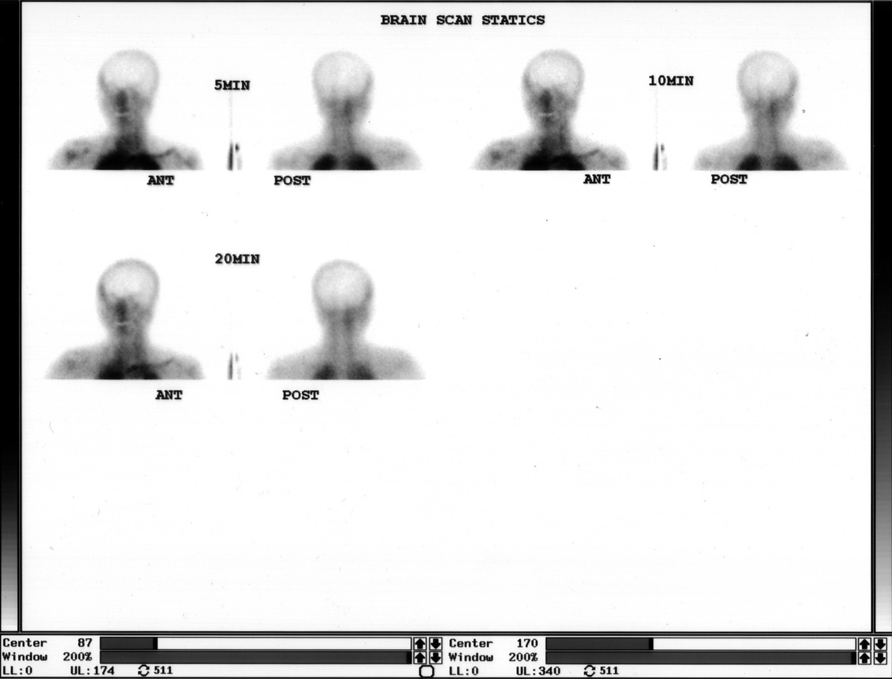 Brain Death Scan: Absent Cerebral Perfusion
Ictal SPECT: Hyperperfusion at the Seizure Focus
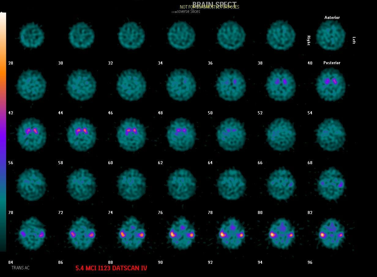 Dementia with Lewy Bodies on DaTscan
Alzheimer’s Disease on FDG PET
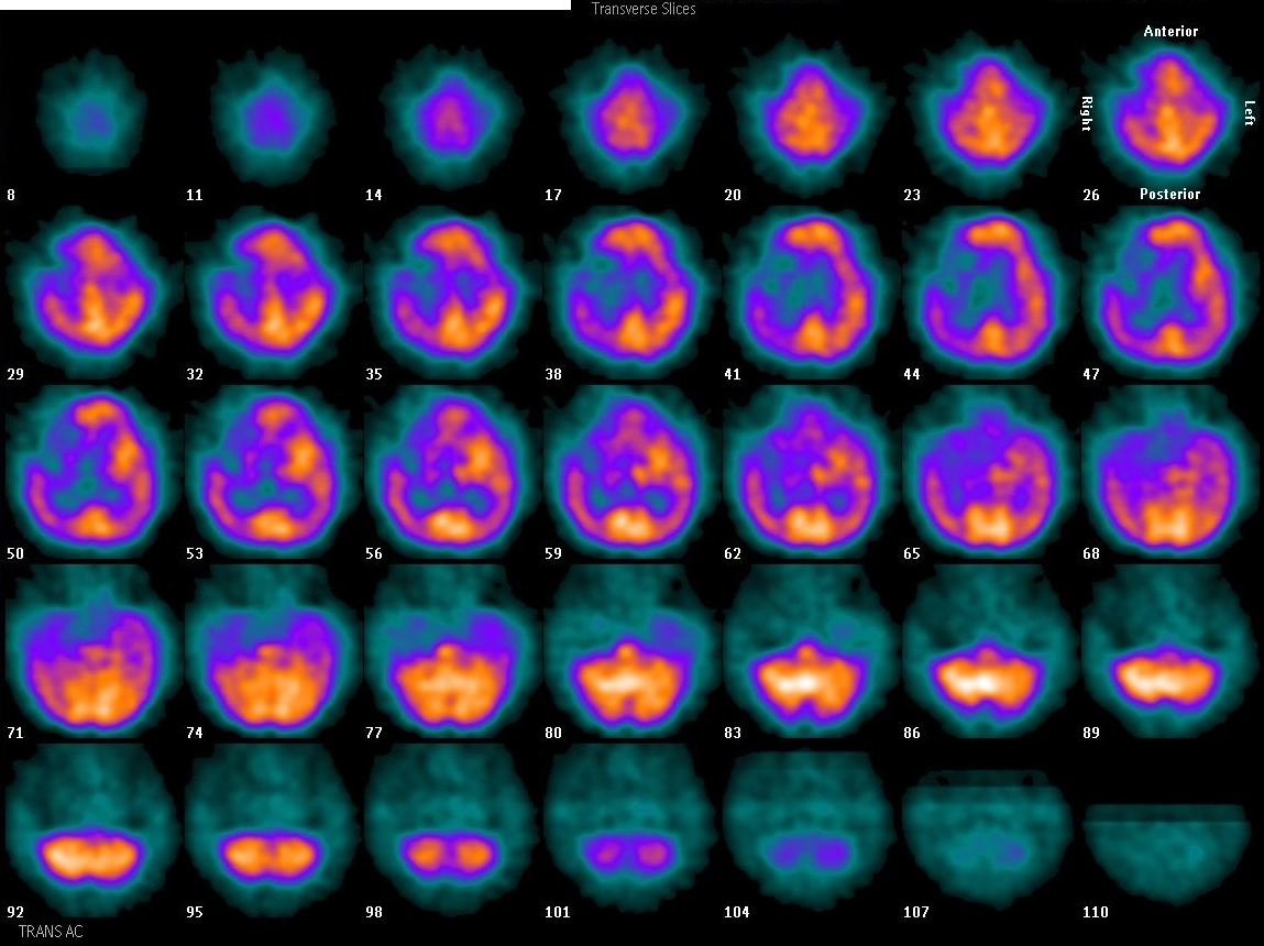 Focal Perfusion Defect on Brain SPECT
Brain Perfusion SPECT: Injection Environment
All 130 nuclear medicine images by category →
Practise image interpretation in the app
All 130 images as an interactive quiz, alongside 500 NMTCB and ARRT(N) questions with worked explanations, case studies and timed mock exams. 25 questions are free, no card and no account.