Alzheimer’s Disease on FDG PET
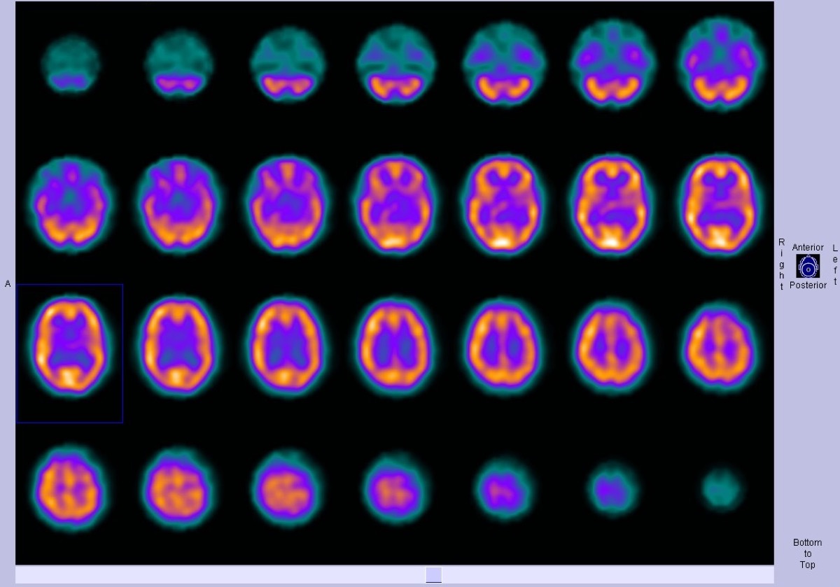
CC BY-SA 3.0 · Dr Bruno Di Muzio / Radiopaedia / Wikimedia Commons · source file
Question
On FDG-PET in early Alzheimer's disease, glucose hypometabolism (decreased FDG uptake) is first characteristically seen in which brain region?
- AFrontal lobes and Broca's area
- BPosterior cingulate cortex and precuneus, followed by parietal and temporal cortex
- CBasal ganglia and thalamus
- DOccipital cortex and primary visual areas
Show the answer and explanation
B. Posterior cingulate cortex and precuneus, followed by parietal and temporal cortex
In early Alzheimer's disease, FDG-PET hypometabolism first appears in the POSTERIOR CINGULATE CORTEX and PRECUNEUS — regions involved in default mode network function and early AD pathology. As AD progresses, hypometabolism spreads to the bilateral inferior parietal cortex and posterior temporal cortex. Primary motor and sensory cortices, basal ganglia, and cerebellum are relatively spared until late stages. This characteristic posterior-predominant pattern on FDG-PET (or perfusion SPECT) helps distinguish AD from frontotemporal dementia (frontal/anterior temporal) and Lewy body dementia (parieto-occipital, with posterior cingulate sparing — the 'cingulate island sign').
More brain imaging images
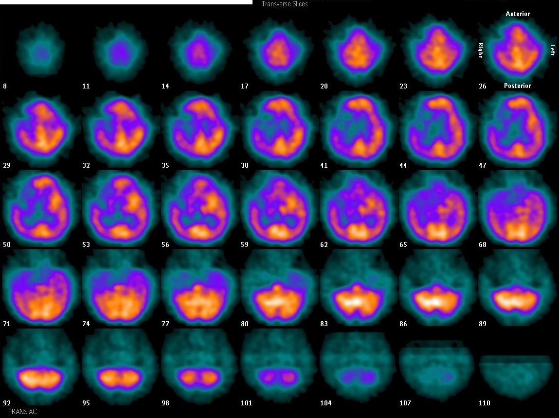 Focal Perfusion Defect on Brain SPECT
Brain Perfusion SPECT: Injection Environment
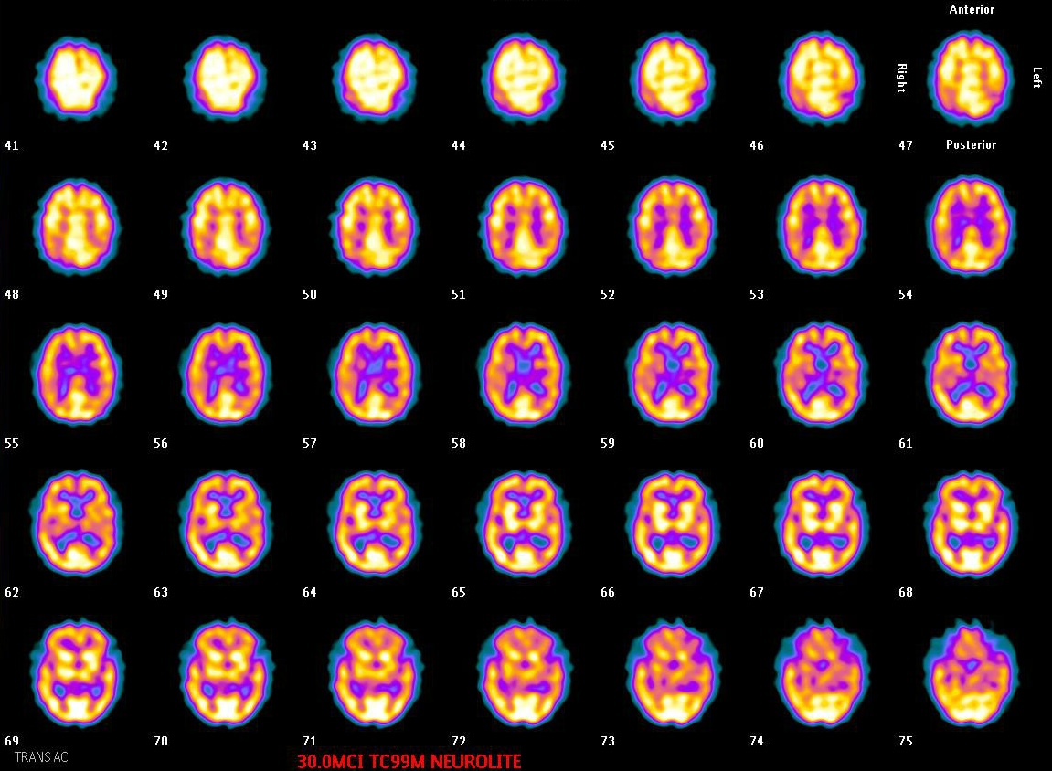 Normal Tc-99m ECD Brain SPECT: Gray vs White Matter
Brain SPECT: When to Image After ECD Injection
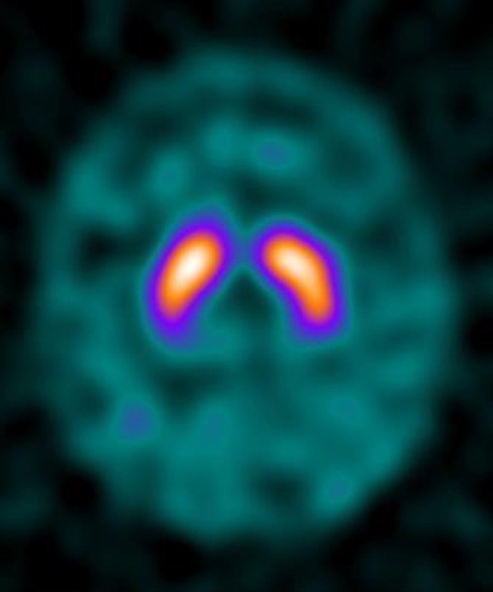 DaTscan (I-123 Ioflupane): What It Evaluates
Normal Brain Perfusion SPECT with Tc-99m HMPAO
All 130 nuclear medicine images by category →
Practise image interpretation in the app
All 130 images as an interactive quiz, alongside 500 NMTCB and ARRT(N) questions with worked explanations, case studies and timed mock exams. 25 questions are free, no card and no account.