Focal Perfusion Defect on Brain SPECT
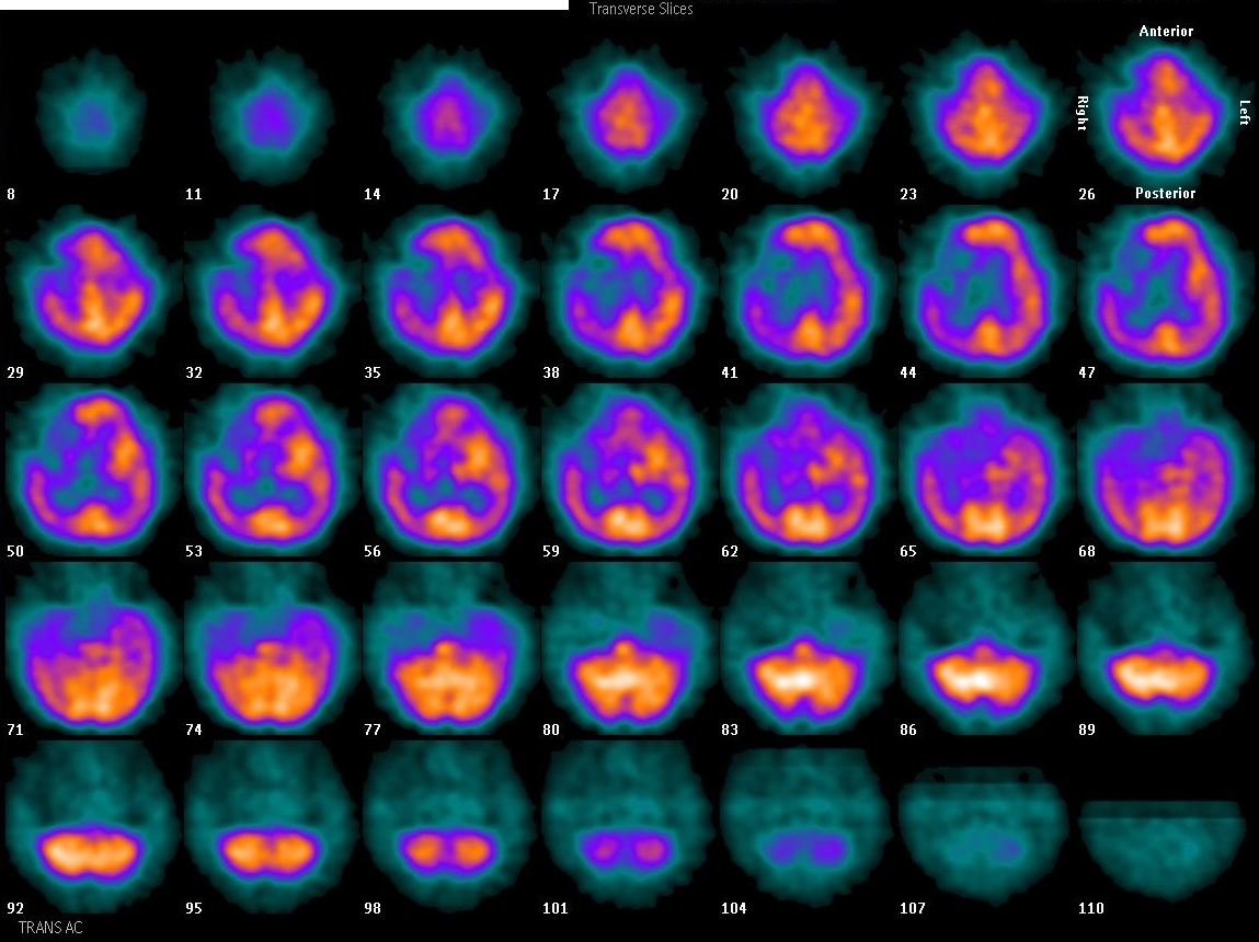
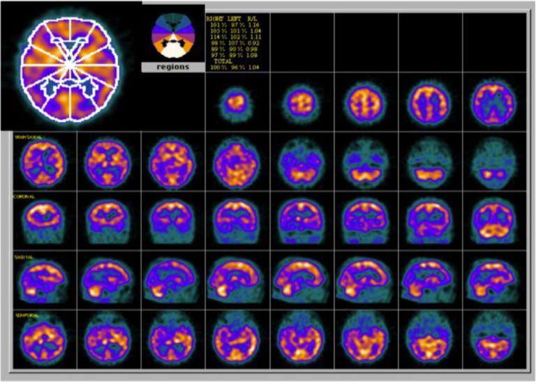
Provided clinical image
Question
This series of transverse brain slices demonstrates a focal region of reduced tracer uptake in one cerebral hemisphere consistent with a vascular territory distribution. Which radiopharmaceutical was most likely used to generate these cerebral perfusion images?
- ATc-99m HMPAO (Ceretec), which crosses the blood-brain barrier and distributes proportional to regional cerebral blood flow
- BTc-99m MDP (medronate), which localizes to hydroxyapatite crystals in areas of active bone remodeling and turnover
- CTc-99m MAA (macroaggregated albumin), which lodges in pulmonary capillaries proportional to regional lung blood flow
- DTc-99m sestamibi (Cardiolite), which accumulates in mitochondria-rich myocardium proportional to coronary blood flow
Show the answer and explanation
A. Tc-99m HMPAO (Ceretec), which crosses the blood-brain barrier and distributes proportional to regional cerebral blood flow
Tc-99m HMPAO (hexamethylpropyleneamine oxime, Ceretec) is a lipophilic radiopharmaceutical that crosses the intact blood-brain barrier and is trapped intracellularly in proportion to regional cerebral blood flow (rCBF). It is one of two main agents used for brain perfusion SPECT, along with Tc-99m ECD (Neurolite). The focal defect seen here in a vascular territory distribution is characteristic of cerebral infarction — an area of absent or severely reduced perfusion resulting from arterial occlusion.
More brain imaging images
Brain Perfusion SPECT: Injection Environment
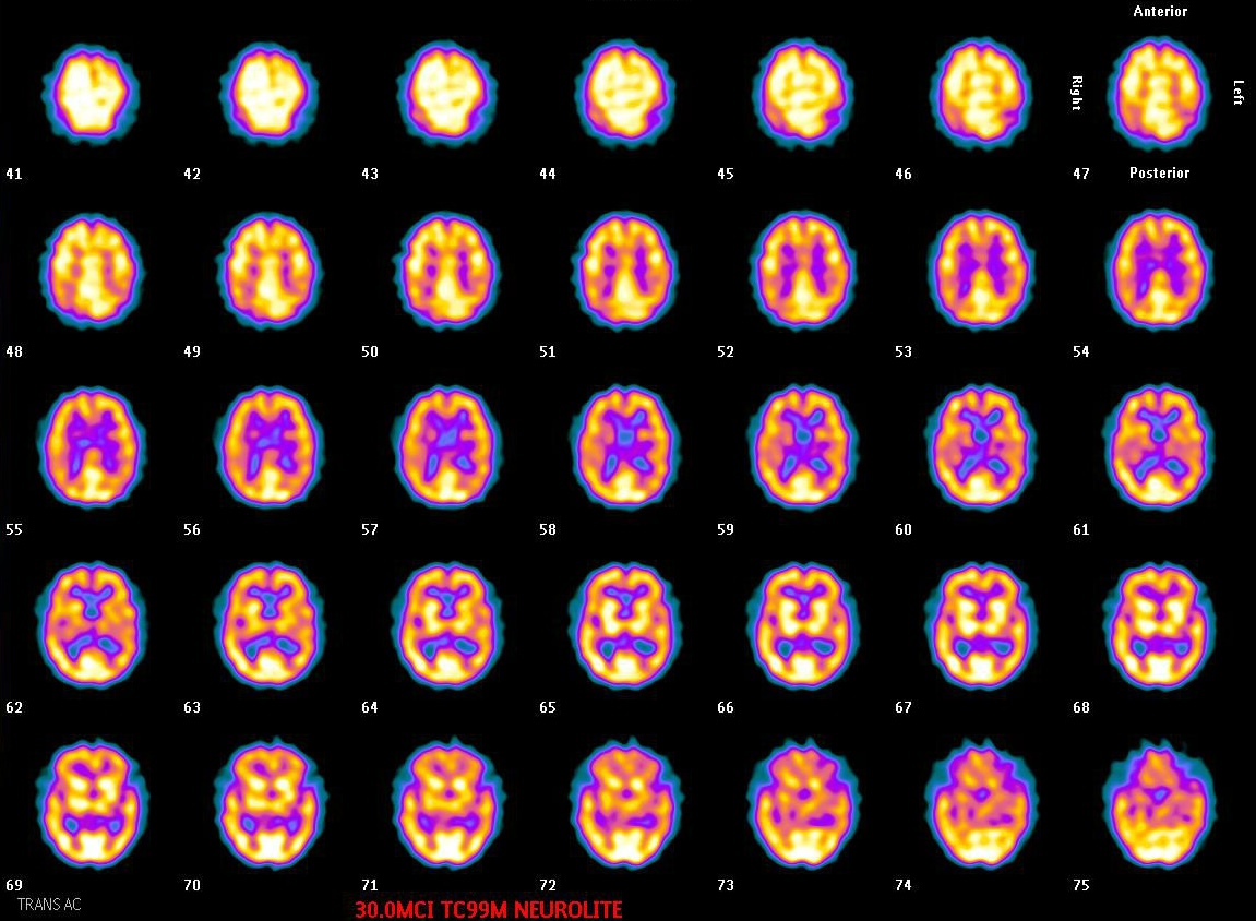 Normal Tc-99m ECD Brain SPECT: Gray vs White Matter
Brain SPECT: When to Image After ECD Injection
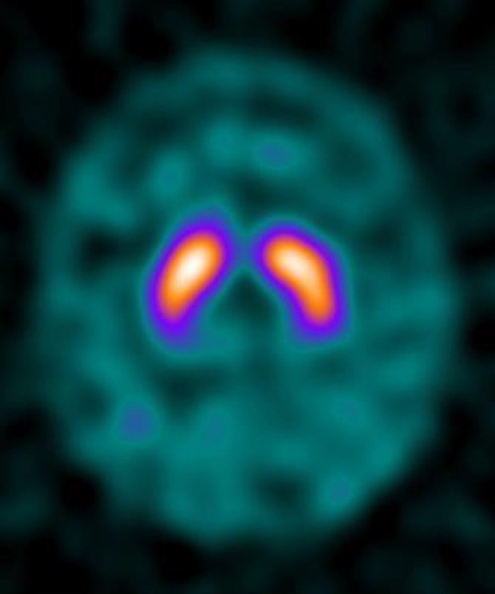 DaTscan (I-123 Ioflupane): What It Evaluates
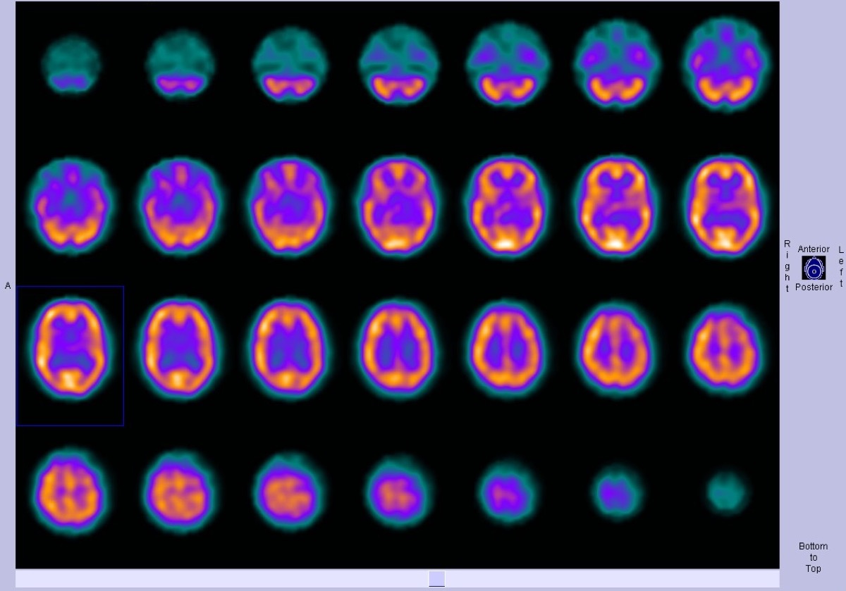 Normal Brain Perfusion SPECT with Tc-99m HMPAO
 Alzheimer’s Disease on Brain Perfusion SPECT
All 130 nuclear medicine images by category →
Practise image interpretation in the app
All 130 images as an interactive quiz, alongside 500 NMTCB and ARRT(N) questions with worked explanations, case studies and timed mock exams. 25 questions are free, no card and no account.