Normal Tc-99m ECD Brain SPECT: Gray vs White Matter
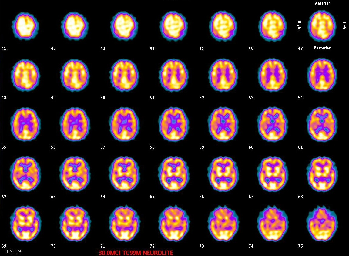
Provided clinical image
Question
These transverse brain slices show symmetric, bilateral tracer distribution throughout the cerebral cortex with a uniform appearance. In a properly acquired normal brain perfusion study, which description best characterizes the expected tracer distribution pattern?
- AWhite matter structures show higher uptake than gray matter cortex due to greater myelinated fiber density
- BGray and white matter display identical uptake levels with no differentiation visible between brain structures
- CGray matter cortex and basal ganglia show higher uptake than white matter, with minimal activity in ventricular regions
- DActivity is confined to deep white matter with cortical gray matter appearing nearly photopenic throughout the study
Show the answer and explanation
C. Gray matter cortex and basal ganglia show higher uptake than white matter, with minimal activity in ventricular regions
In a normal brain perfusion SPECT, gray matter structures — including the cerebral cortex, basal ganglia, thalami, and cerebellum — show significantly higher tracer uptake compared to white matter because they have greater metabolic demand and blood flow per unit volume. The ventricular regions appear as areas of very low or absent activity. Clear gray-white matter differentiation is also an important quality indicator: poor differentiation may suggest technical issues, sedation effects, or generalized hypoperfusion.
More brain imaging images
Brain SPECT: When to Image After ECD Injection
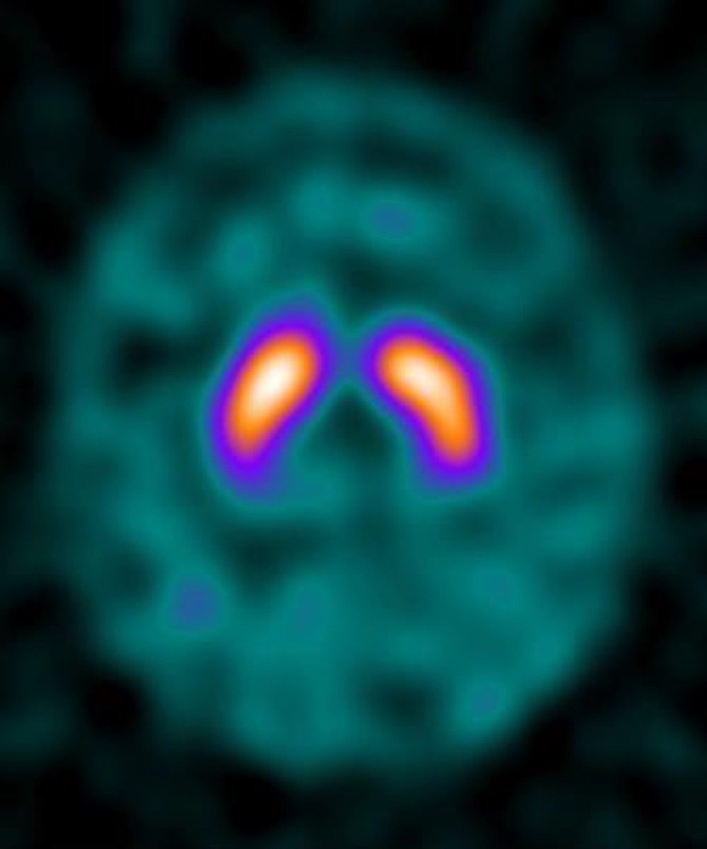 DaTscan (I-123 Ioflupane): What It Evaluates
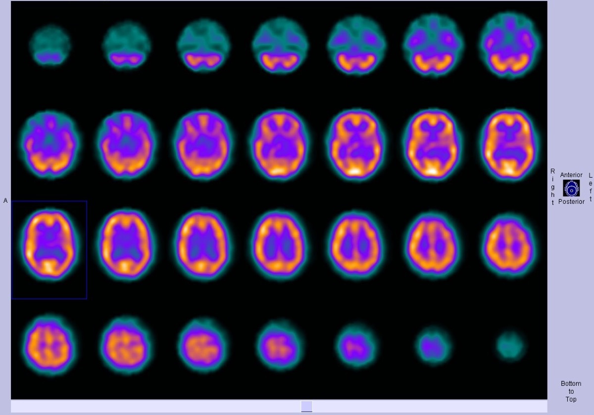 Normal Brain Perfusion SPECT with Tc-99m HMPAO
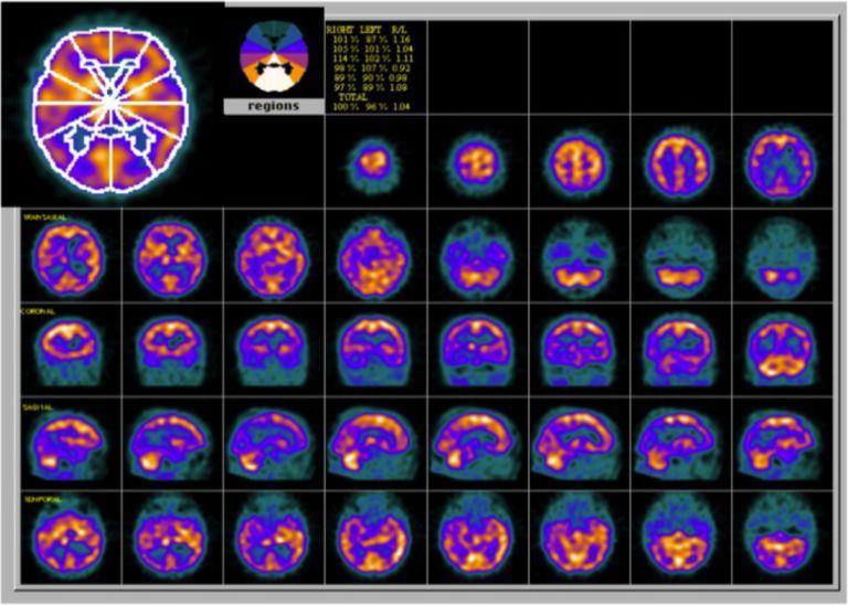 Alzheimer’s Disease on Brain Perfusion SPECT
Parkinson’s Disease on DaTscan: Loss of the Comma Shape
Frontotemporal Dementia on Brain Perfusion SPECT
All 130 nuclear medicine images by category →
Practise image interpretation in the app
All 130 images as an interactive quiz, alongside 500 NMTCB and ARRT(N) questions with worked explanations, case studies and timed mock exams. 25 questions are free, no card and no account.