Brain Perfusion SPECT: Injection Environment
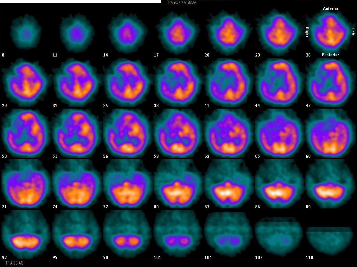
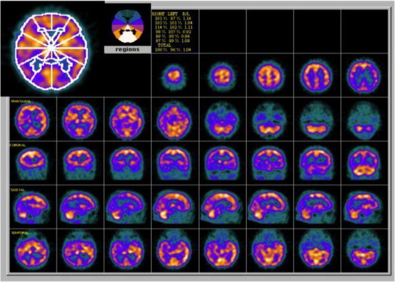
Provided clinical image
Question
When performing a brain perfusion study like the one shown, what environment should be prepared for the patient immediately before and during radiopharmaceutical injection to ensure accurate mapping of baseline cerebral blood flow?
- AA brightly lit procedure room with standard background noise and normal patient conversation allowed during injection
- BA quiet, dimly lit room with eyes open and minimal auditory and visual stimulation before and throughout injection
- CA darkened room with the patient instructed to keep both eyes tightly closed throughout the entire uptake period
- DA standard waiting area where the patient may watch television or use their phone until transfer to the camera room
Show the answer and explanation
B. A quiet, dimly lit room with eyes open and minimal auditory and visual stimulation before and throughout injection
Brain perfusion agents such as Tc-99m HMPAO and Tc-99m ECD fix rapidly in the brain in proportion to cerebral blood flow at the time of injection. Because sensory stimulation (light, sound, movement) increases regional blood flow to corresponding cortical areas, the patient must rest in a quiet, dimly lit room with minimal stimulation for at least 10–20 minutes before and during injection. Eyes should remain open — not closed — because a closed visual cortex maintains baseline activity, while eyes open but in dim light avoids driving occipital cortex uptake.
More brain imaging images
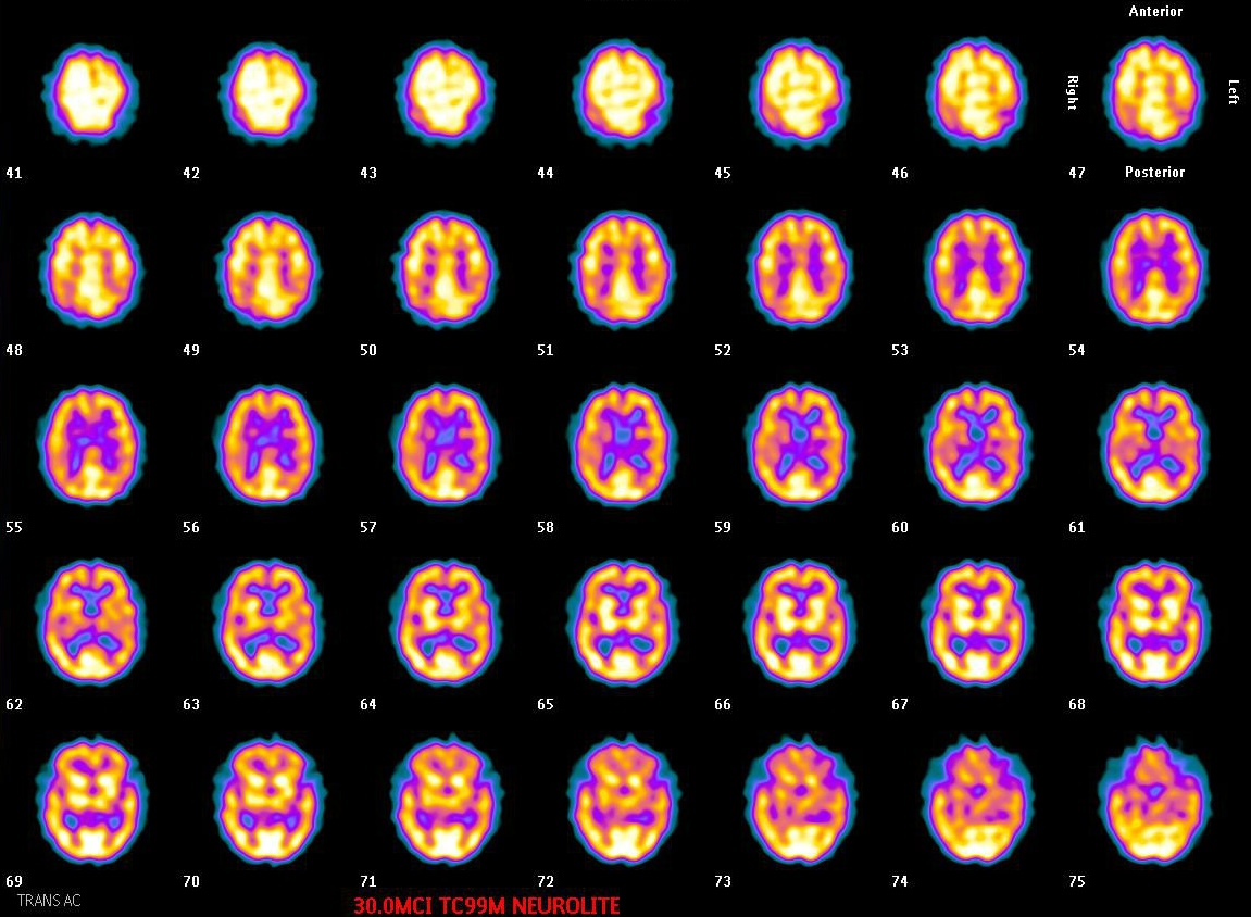 Normal Tc-99m ECD Brain SPECT: Gray vs White Matter
Brain SPECT: When to Image After ECD Injection
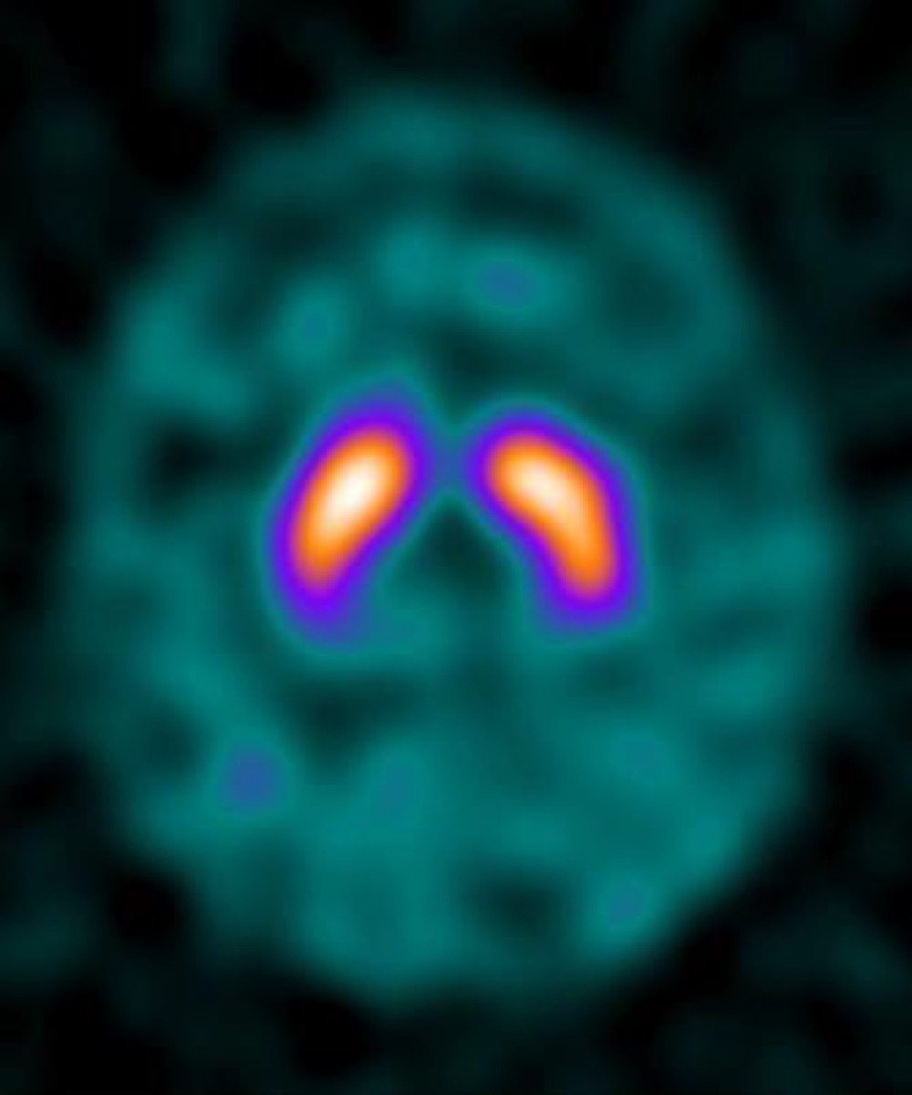 DaTscan (I-123 Ioflupane): What It Evaluates
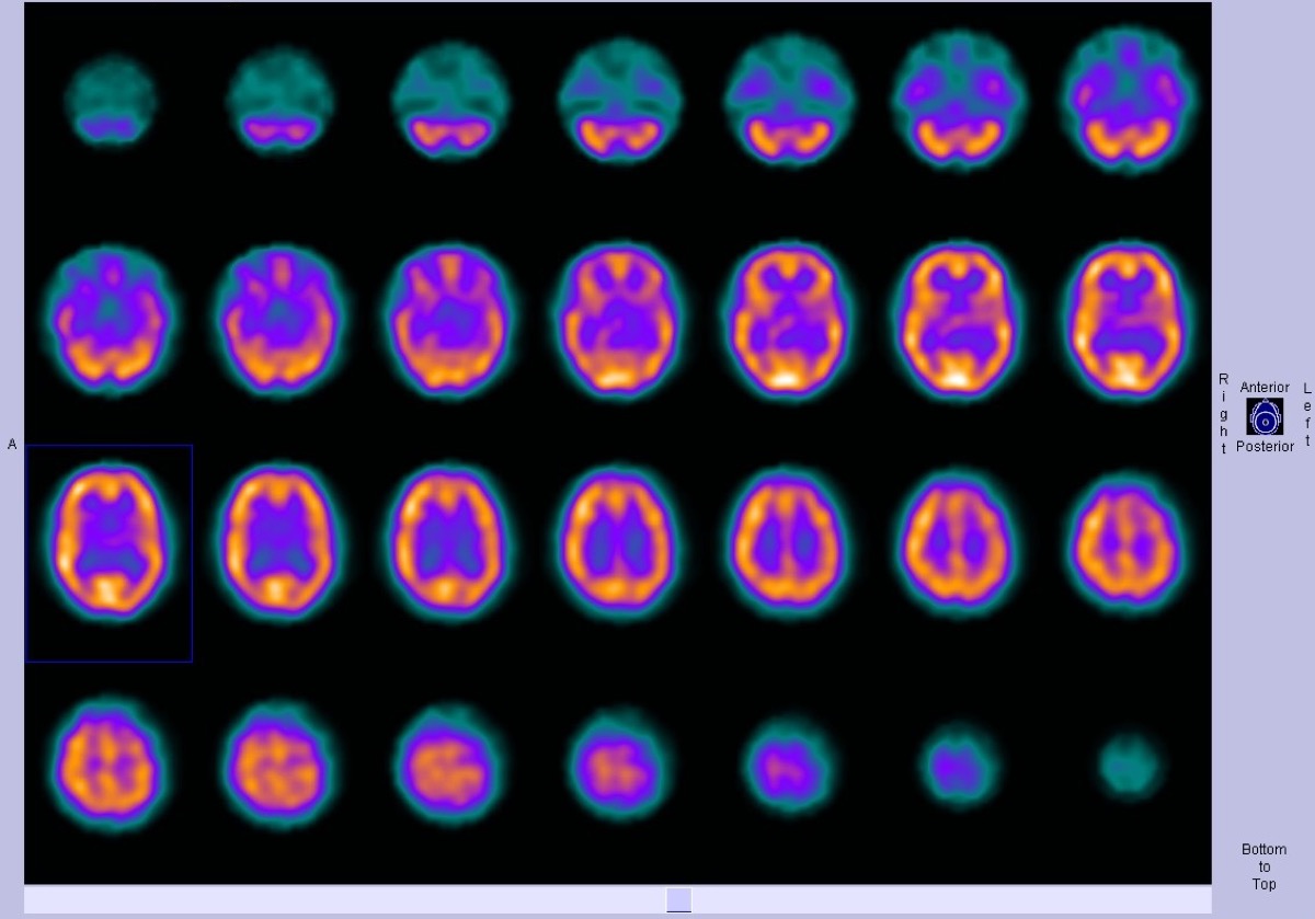 Normal Brain Perfusion SPECT with Tc-99m HMPAO
 Alzheimer’s Disease on Brain Perfusion SPECT
Parkinson’s Disease on DaTscan: Loss of the Comma Shape
All 130 nuclear medicine images by category →
Practise image interpretation in the app
All 130 images as an interactive quiz, alongside 500 NMTCB and ARRT(N) questions with worked explanations, case studies and timed mock exams. 25 questions are free, no card and no account.