Brain SPECT: When to Image After ECD Injection
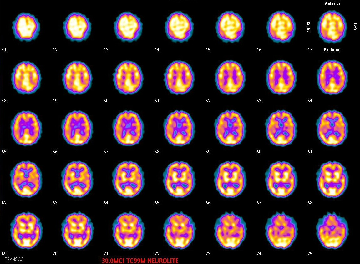
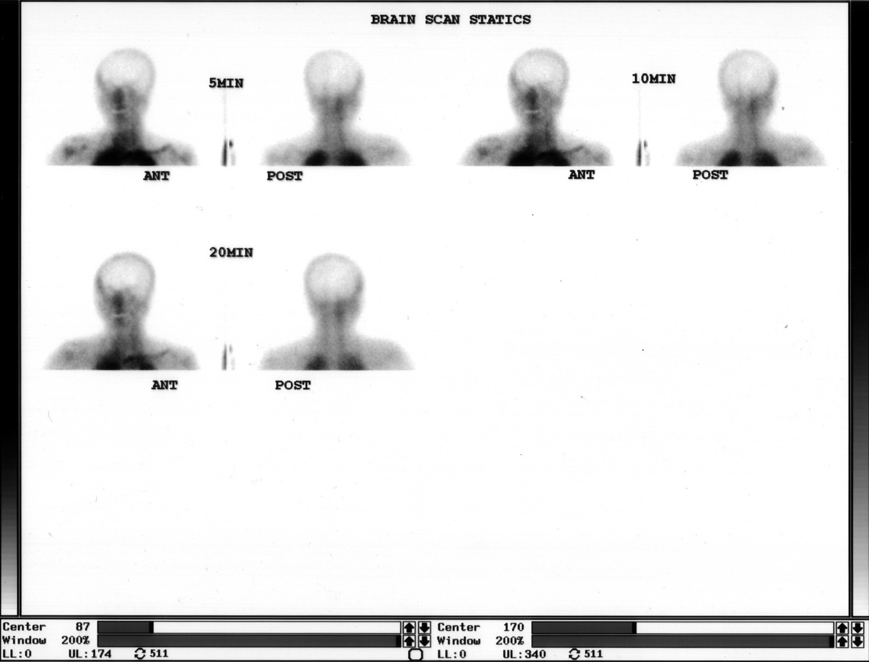
Provided clinical image
Question
In the type of brain perfusion study shown here, once the radiopharmaceutical has been injected in the appropriate resting environment, approximately how long after injection should SPECT image acquisition typically begin?
- AWithin 60 seconds, before the tracer begins to redistribute away from its initial perfusion pattern in the brain
- B6–24 hours after injection to allow complete clearance of non-specific circulating background activity
- CImmediately after injection while the patient remains in the quiet supine position to capture peak brain activity
- D15–30 minutes after injection, allowing the tracer to fix in brain tissue and background activity to clear sufficiently
Show the answer and explanation
D. 15–30 minutes after injection, allowing the tracer to fix in brain tissue and background activity to clear sufficiently
For Tc-99m HMPAO and Tc-99m ECD brain perfusion studies, SPECT acquisition typically begins 15–30 minutes after injection. By this time, the lipophilic tracer has crossed the blood-brain barrier, undergone intracellular conversion to a hydrophilic form, and is fixed in brain parenchyma proportional to perfusion at the time of injection. Starting acquisition too early captures residual circulating tracer and elevated background; waiting too long — especially with Tc-99m HMPAO — may reduce image quality as brain-to-background ratios decline over time.
More brain imaging images
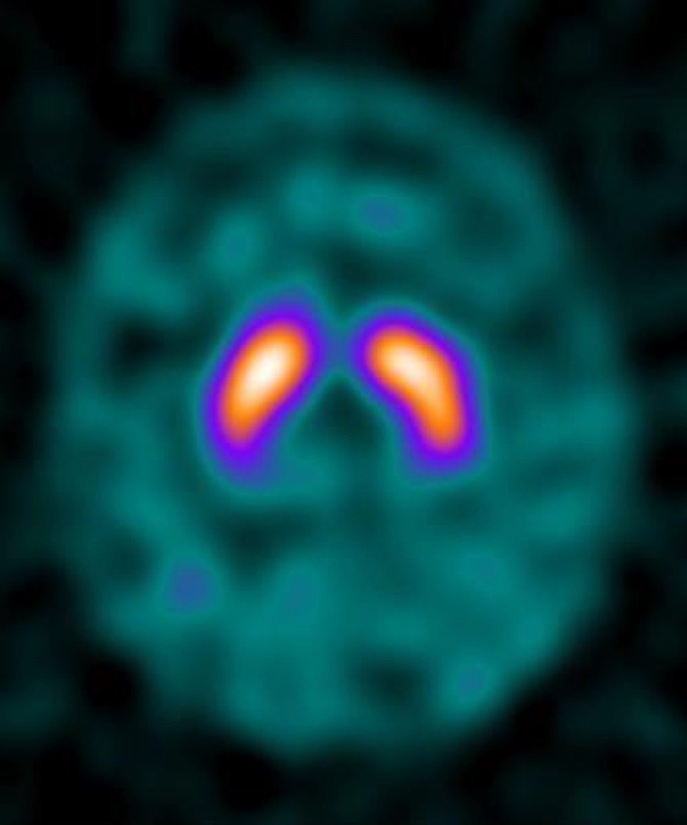 DaTscan (I-123 Ioflupane): What It Evaluates
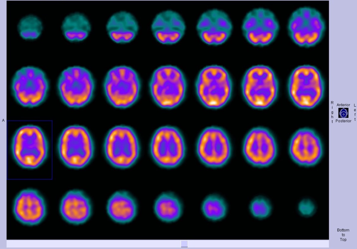 Normal Brain Perfusion SPECT with Tc-99m HMPAO
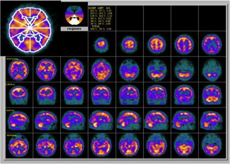 Alzheimer’s Disease on Brain Perfusion SPECT
Parkinson’s Disease on DaTscan: Loss of the Comma Shape
Frontotemporal Dementia on Brain Perfusion SPECT
 Brain Death Scan: Absent Cerebral Perfusion
All 130 nuclear medicine images by category →
Practise image interpretation in the app
All 130 images as an interactive quiz, alongside 500 NMTCB and ARRT(N) questions with worked explanations, case studies and timed mock exams. 25 questions are free, no card and no account.