FDG-Avid Benign Disease: Infection and Inflammation Mimics
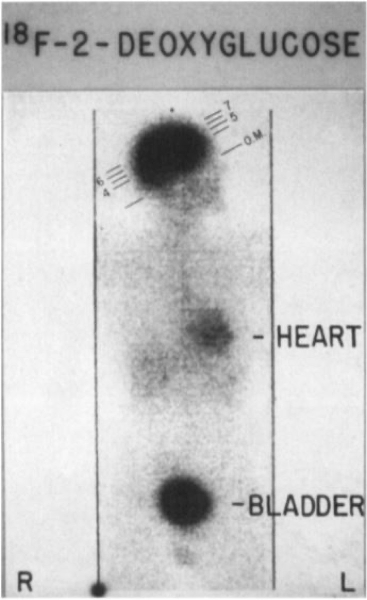
CC BY-SA 4.0 · Pennradiology / Wikimedia Commons · source file
Question
A patient with a history of non-Hodgkin lymphoma has an FDG PET/CT showing an intensely FDG-avid mediastinal mass. Biopsy reveals granulomatous inflammation (sarcoidosis), not lymphoma recurrence. This scenario illustrates an important FDG PET pitfall — which of the following represents a common cause of FALSE-POSITIVE FDG uptake in oncologic PET?
- ANormal variant brain uptake
- BRenal excretion of FDG
- CInfection and inflammation (including sarcoidosis, tuberculosis, fungal infections, post-surgical healing) — inflammatory cells are highly FDG-avid
- DCardiac motion artifact
Show the answer and explanation
C. Infection and inflammation (including sarcoidosis, tuberculosis, fungal infections, post-surgical healing) — inflammatory cells are highly FDG-avid
FDG (fluorodeoxyglucose) is a glucose analogue taken up by any metabolically active cell — not just cancer cells. Activated macrophages, neutrophils, and other inflammatory cells have dramatically upregulated glucose metabolism and can show very intense FDG uptake indistinguishable from malignancy. Common causes of false-positive FDG uptake include: sarcoidosis (classic pitfall in lymphoma); granulomatous infections (TB, histoplasmosis); post-surgical/radiation inflammation; brown fat (supraclavicular/mediastinal); healing fractures; inflammatory bowel disease. Correlating with CT morphology, clinical history, and if needed biopsy is essential to avoid misinterpreting FDG-avid inflammation as tumor recurrence.
More pet and oncology images
How SUV Is Calculated on FDG PET
FDG PET/CT Staging of Non-Small Cell Lung Cancer
Brown Fat Uptake on FDG PET
F-18 Half-Life and Decay Mode
PET Coincidence Detection of 511 keV Photons
 MIBG Scan for Neuroblastoma Staging
All 130 nuclear medicine images by category →
Practise image interpretation in the app
All 130 images as an interactive quiz, alongside 500 NMTCB and ARRT(N) questions with worked explanations, case studies and timed mock exams. 25 questions are free, no card and no account.