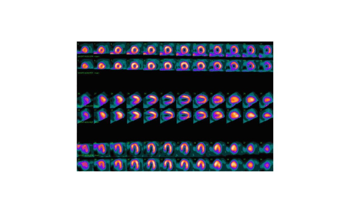
LAD Territory Perfusion Defect on SPECT
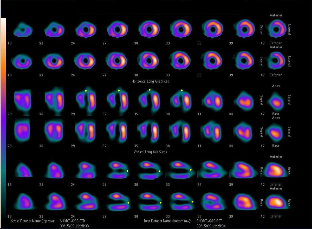
CC BY-SA 3.0 · Wikimedia Commons · source file
Question
On myocardial perfusion SPECT, a perfusion defect involving the anterior wall, anteroseptum, and apex is present at stress and partially fills in at rest (partially reversible). This territory is supplied by the left anterior descending artery (LAD). An LAD territory perfusion defect indicates which vessel is affected?
- AThe LAD territory (anterior wall, anteroseptum, and apex) is the most common site of MI and predicts significant CAD in the LAD distribution
- BRCA territory
- CPosterior descending artery (PDA) territory
- DLCX territory isolated to the lateral wall
Show the answer and explanation
A. The LAD territory (anterior wall, anteroseptum, and apex) is the most common site of MI and predicts significant CAD in the LAD distribution
The left anterior descending artery (LAD) supplies the anterior wall, anterior septum, and cardiac apex (and part of the lateral wall via diagonal branches). An LAD territory perfusion defect is the most common and highest-risk finding on MPI because LAD occlusion (the 'widow maker') threatens a large myocardial territory. A partially reversible defect indicates ischemia with some viable myocardium in the LAD territory. The LAD territory encompasses approximately 45–50% of the total left ventricular myocardium, making LAD disease the highest-risk anatomic pattern.
More cardiac imaging images
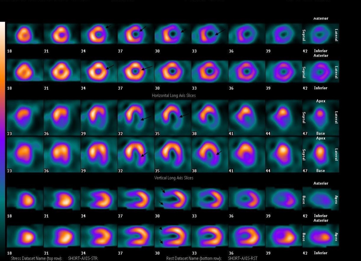 Multivessel Coronary Disease on Stress MPI
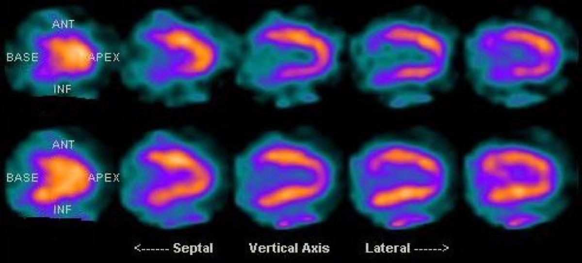 Prone Imaging in Cardiac SPECT
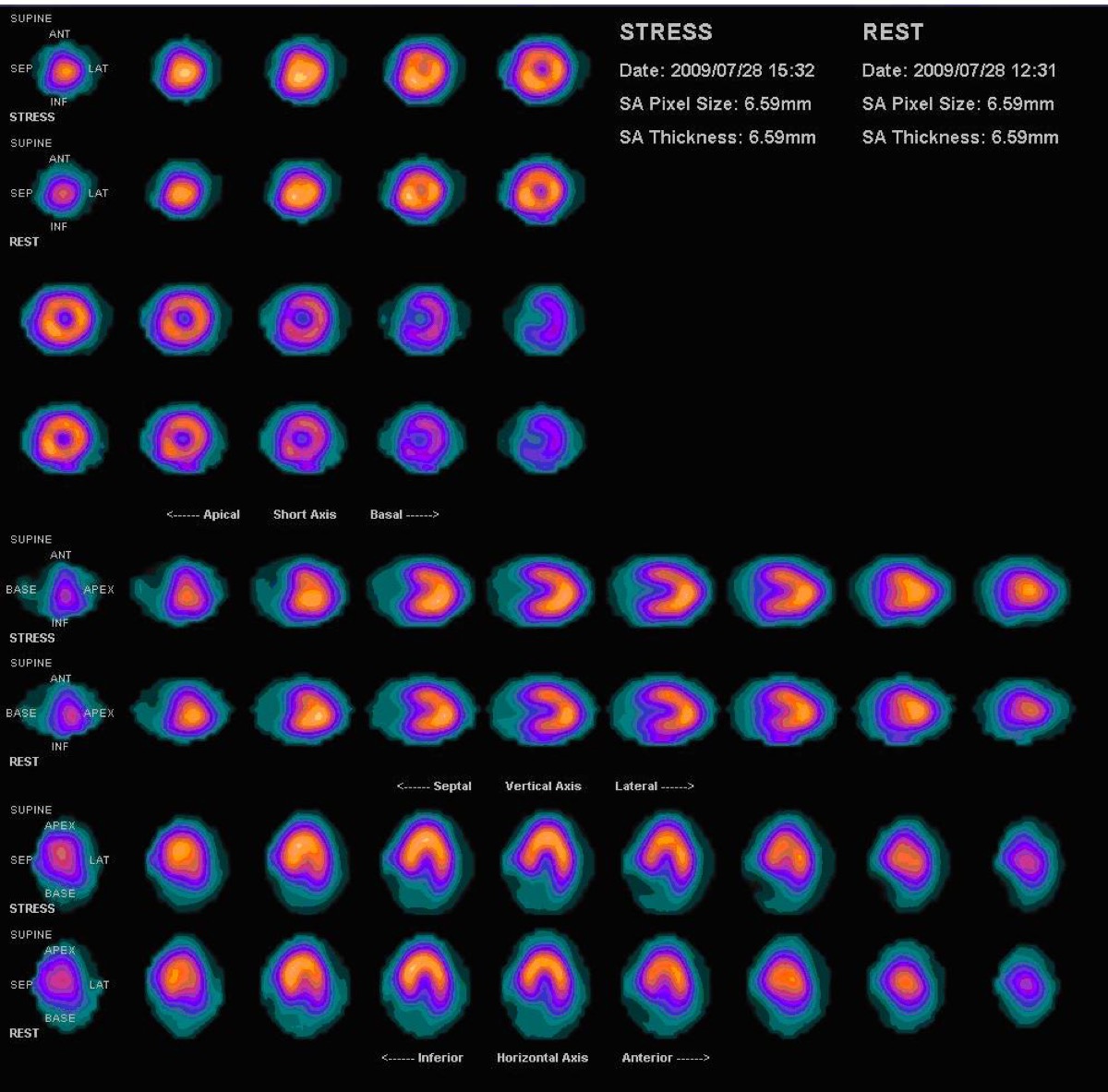 Prominent Right Ventricle on MPI: RV Hypertrophy
 Dobutamine Stress for Myocardial Perfusion Imaging
PET vs SPECT MPI: Absolute Myocardial Blood Flow
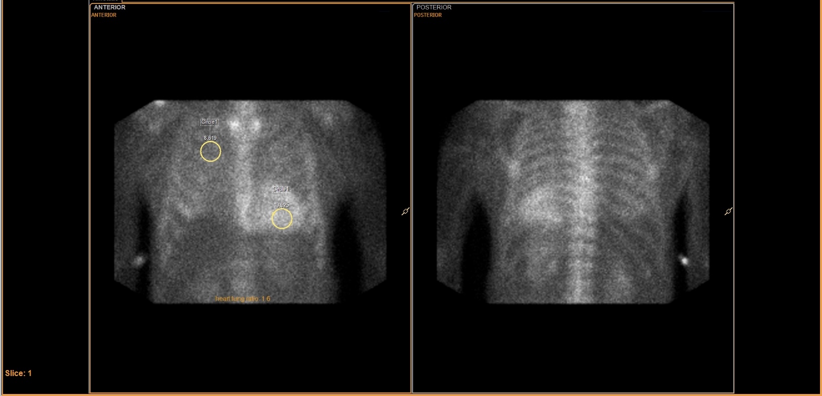 Tc-99m PYP Heart-to-Lung Ratio for Cardiac Amyloidosis
All 130 nuclear medicine images by category →
Practise image interpretation in the app
All 130 images as an interactive quiz, alongside 500 NMTCB and ARRT(N) questions with worked explanations, case studies and timed mock exams. 25 questions are free, no card and no account.