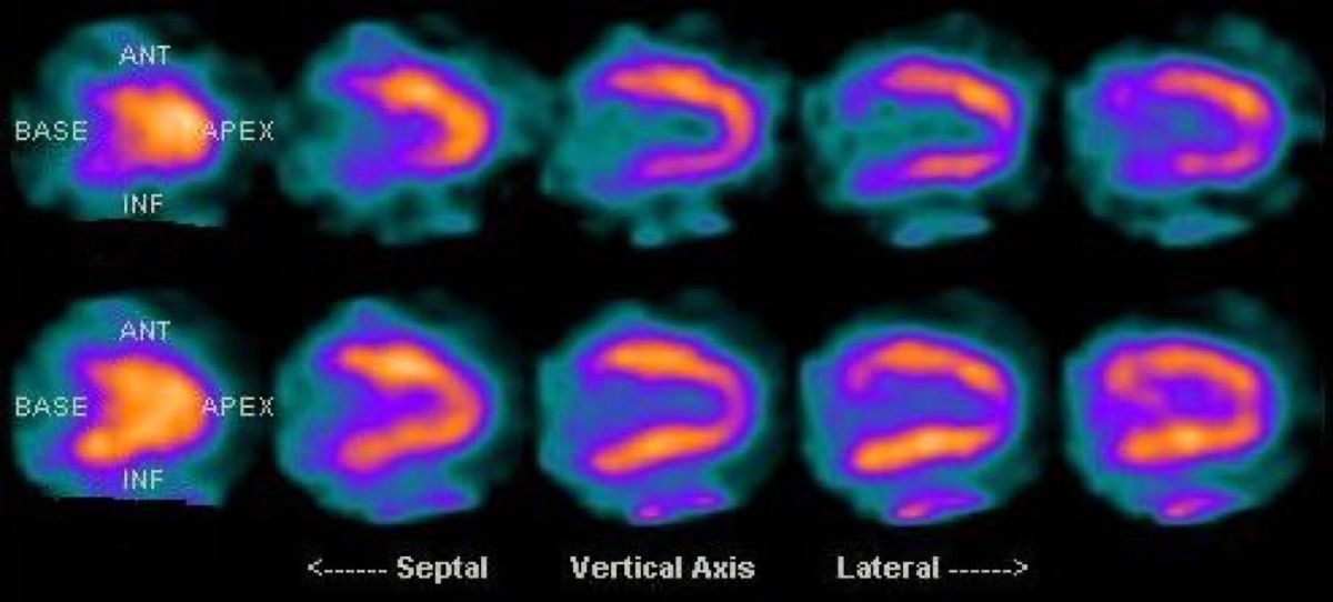
Inferior Wall Ischemia: Right Coronary Artery Territory
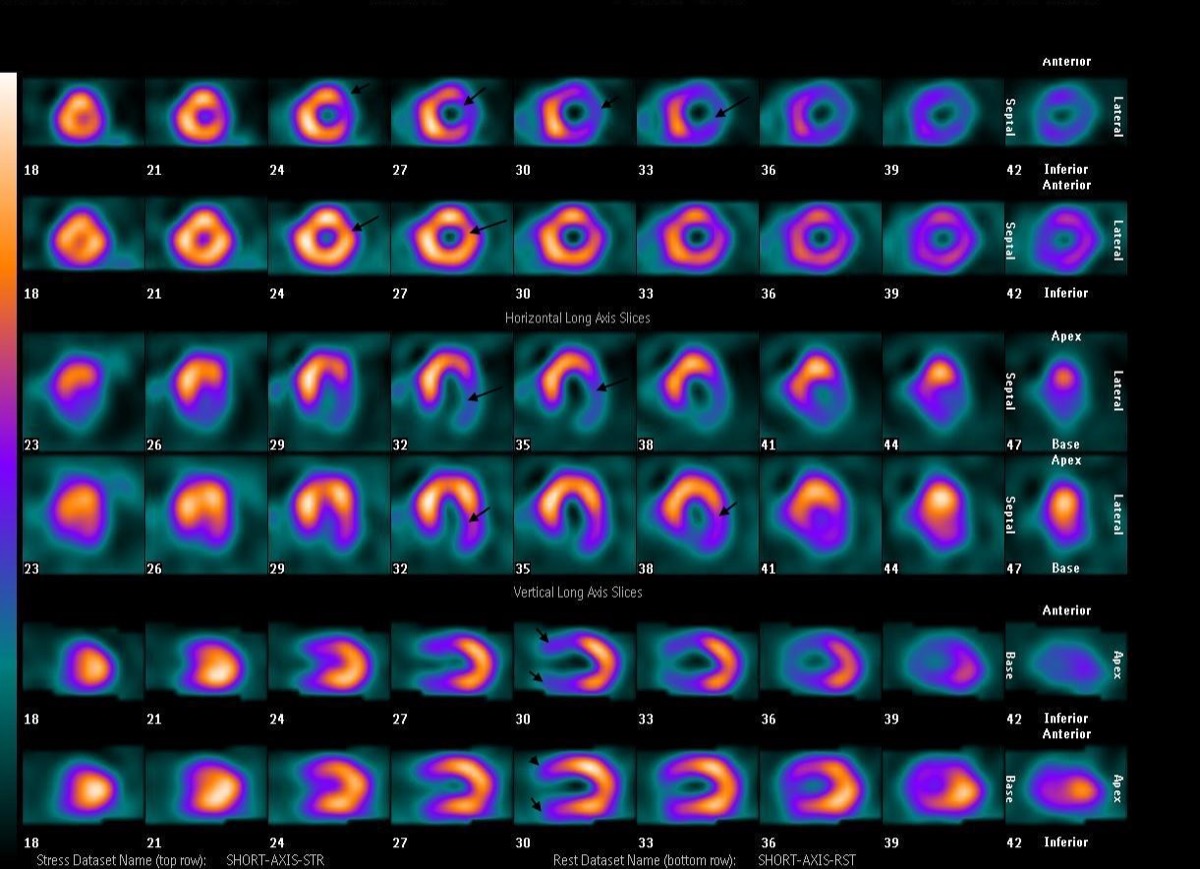
CC BY-SA 4.0 · Wikimedia Commons · source file
Question
A reversible (stress-induced) inferior wall perfusion defect is identified on MPI. The inferior and inferolateral walls of the left ventricle are supplied primarily by which coronary artery in a right-dominant coronary system?
- ALeft anterior descending artery (LAD)
- BRight coronary artery (RCA) — supplies the inferior wall via the posterior descending artery
- CLeft circumflex artery (LCX)
- DLeft main coronary artery
Show the answer and explanation
B. Right coronary artery (RCA) — supplies the inferior wall via the posterior descending artery
In a right-dominant coronary system (present in ~85% of individuals), the right coronary artery (RCA) gives rise to the posterior descending artery (PDA), which supplies the inferior wall of the left ventricle, the posterior interventricular septum, and the inferior septum. A reversible inferior wall defect on MPI therefore indicates RCA territory ischemia. In a left-dominant system, the LCX gives rise to the PDA. The inferior wall is the most common territory for attenuation artifact (diaphragm in men, inferior portion in women), so gated SPECT and prone imaging help distinguish artifact from true RCA ischemia.
More cardiac imaging images
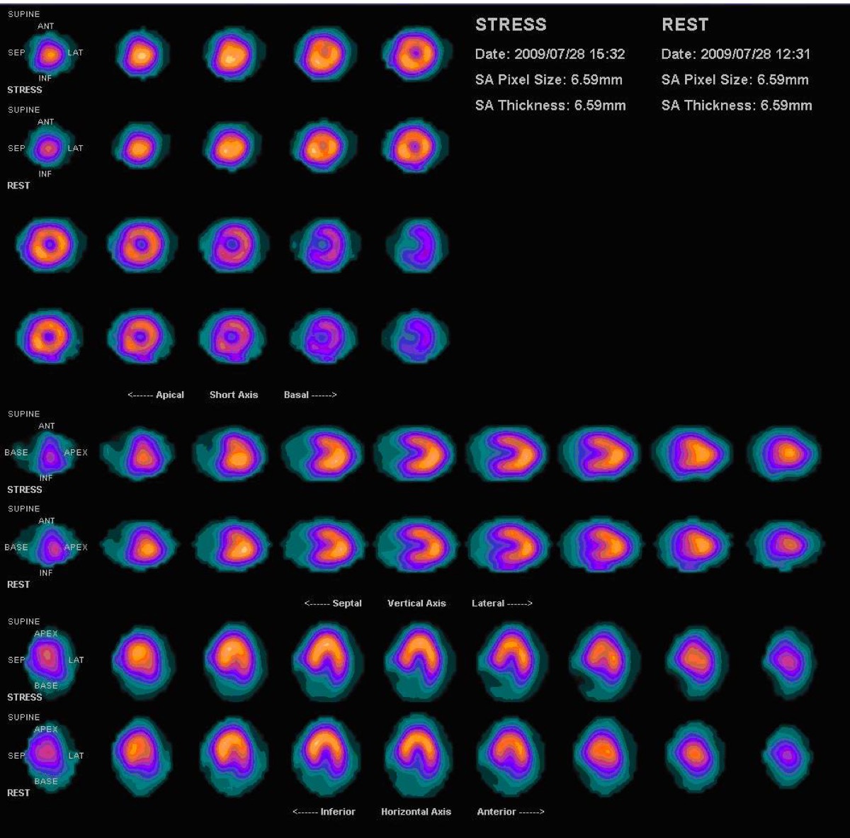 Vasodilator Stress Contraindications: Caffeine and Bronchospasm
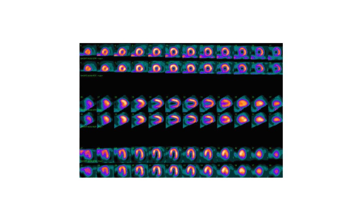 Gated SPECT: Ejection Fraction and Ventricular Volumes
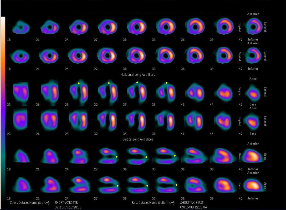 LAD Territory Perfusion Defect on SPECT
Multivessel Coronary Disease on Stress MPI
 Prone Imaging in Cardiac SPECT
Prominent Right Ventricle on MPI: RV Hypertrophy
All 130 nuclear medicine images by category →
Practise image interpretation in the app
All 130 images as an interactive quiz, alongside 500 NMTCB and ARRT(N) questions with worked explanations, case studies and timed mock exams. 25 questions are free, no card and no account.