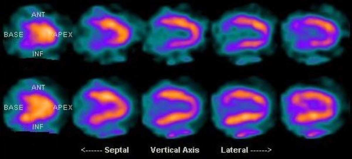
FDG PET Viability: Hibernating Myocardium
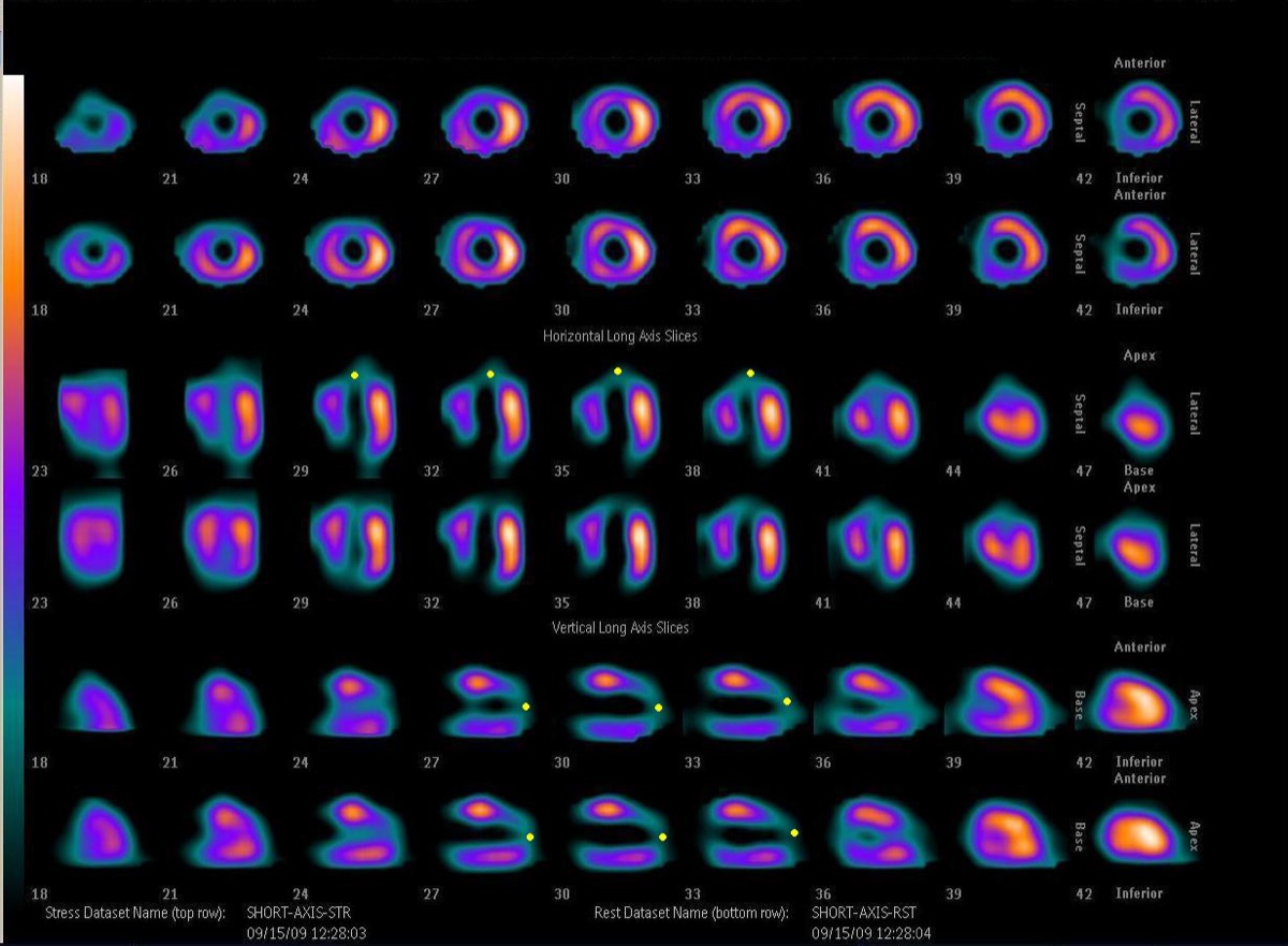
CC BY-SA 3.0 · Wikimedia Commons · source file
Question
A patient has a fixed (non-reversible) anterior wall perfusion defect on MPI suggesting prior LAD infarction. To determine if the infarcted myocardium contains any viable (hibernating) tissue, which additional study would be MOST useful?
- ARepeat pharmacologic stress test
- BExercise electrocardiography
- CFDG PET viability study — hibernating myocardium shows preserved FDG uptake despite reduced perfusion
- DCoronary CTA
Show the answer and explanation
C. FDG PET viability study — hibernating myocardium shows preserved FDG uptake despite reduced perfusion
Hibernating myocardium refers to chronically hypoperfused but viable myocardium that shows wall motion abnormality at rest but can recover function after revascularization. On MPI, it appears as a fixed (non-reversible) perfusion defect — similar to scar. FDG PET viability imaging exploits the fact that viable myocardium switches to glucose metabolism under ischemic conditions: hibernating tissue shows a 'perfusion-metabolism mismatch' (reduced perfusion, preserved FDG uptake). Pure scar shows concordantly reduced perfusion AND reduced FDG uptake. Identifying hibernation guides the decision for revascularization.
More cardiac imaging images
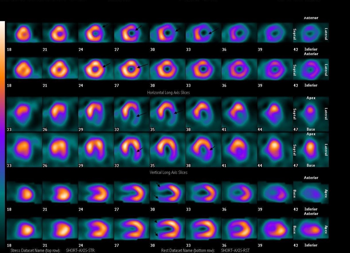 Inferior Wall Ischemia: Right Coronary Artery Territory
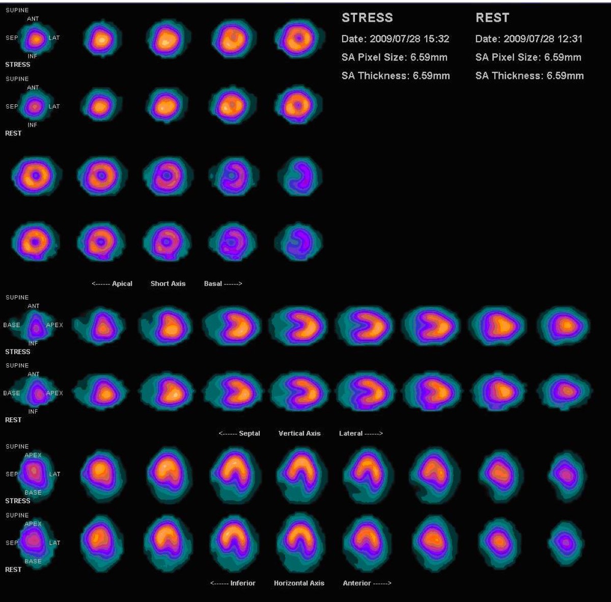 Vasodilator Stress Contraindications: Caffeine and Bronchospasm
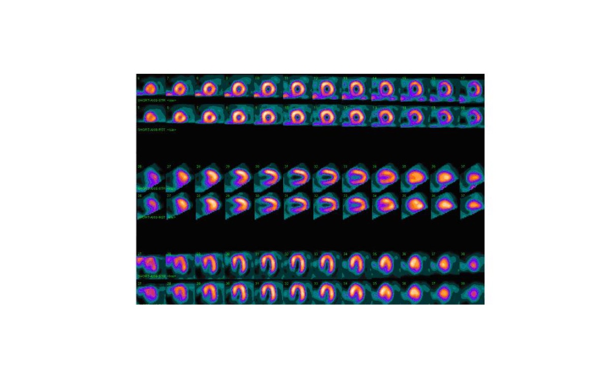 Gated SPECT: Ejection Fraction and Ventricular Volumes
LAD Territory Perfusion Defect on SPECT
Multivessel Coronary Disease on Stress MPI
 Prone Imaging in Cardiac SPECT
All 130 nuclear medicine images by category →
Practise image interpretation in the app
All 130 images as an interactive quiz, alongside 500 NMTCB and ARRT(N) questions with worked explanations, case studies and timed mock exams. 25 questions are free, no card and no account.